File:Fetal cardiac ultrasound 01.jpg

Original file (900 × 600 pixels, file size: 88 KB, MIME type: image/jpeg)
Fetal Cardiac Ultrasound
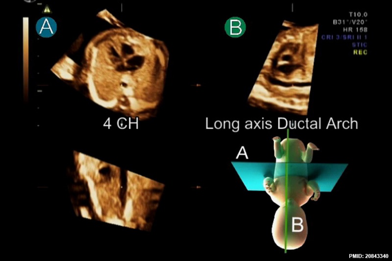
After standardization of fetal position in the STIC volume dataset to place the fetus in the exact dorsal supine position, navigating systematically in the volume usually provides a reproducible image from a corresponded movement. Placing the reference dot in the center of the aorta in the four-chamber (4 CH) view in plane A simultaneously displays long axis of the ductal arch in plane B.
Reference
<pubmed>20843340</pubmed>| Cardiovasc Ultrasound.
Jantarasaengaram and Vairojanavong Cardiovascular Ultrasound 2010 8:41
1476-7120-8-41-3.jpg
doi:10.1186/1476-7120-8-41
File history
Click on a date/time to view the file as it appeared at that time.
| Date/Time | Thumbnail | Dimensions | User | Comment | |
|---|---|---|---|---|---|
| current | 11:53, 10 May 2015 |  | 900 × 600 (88 KB) | Z8600021 (talk | contribs) | After standardization of fetal position in the STIC volume dataset to place the fetus in the exact dorsal supine position, navigating systematically in the volume usually provides a reproducible image from a corresponded movement. Placing the reference... |
You cannot overwrite this file.
File usage
There are no pages that use this file.