File:Fetal Skeletal Muscle Progenitors.png

Original file (1,345 × 1,107 pixels, file size: 1.29 MB, MIME type: image/png)
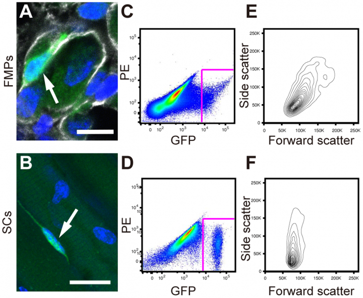
FMPs are more heterogeneous then SCs; Immunohistochemistry of a FMP on longitudinal sections of limbs at E16.5 for GFP (green), Laminin (white). Nuclei were stained with DAPI. An arrow indicates a GFP-positive FMP. Scale bar = 10 µm. (B) Adult myofibers (nuclei were stained with DAPI) isolated from the diaphragms of the Pax3GFP/+ line. An arrow indicates a GFP-positive SC. Scale bar = 25 µm. (C,D) Representative fluorescence-activated cell sorting profiles for (Pax3)GFP+ cells from fetuses (C) and adult muscle (D). (E,F) Forward scatter and side scatter profiles of (Pax3)GFP cells gated in (C) and (D). FMPs, fetal skeletal muscle progenitors; SCs, satellite cells.
Reference;
Citation: Sakai H, Sato T, Sakurai H, Yamamoto T, Hanaoka K, et al. (2013) Fetal Skeletal Muscle Progenitors Have Regenerative Capacity after Intramuscular Engraftment in Dystrophin Deficient Mice. PLoS ONE 8(5): e63016. doi:10.1371/journal.pone.0063016
Copyright: © 2013 Sakai et al. This is an open-access article distributed under the terms of the Creative Commons Attribution License, which permits unrestricted use, distribution, and reproduction in any medium, provided the original author and source are credited.
--Mark Hill Good image uploaded correctly. Reference link is not formatted. Help:Reference_Tutorial FYI I have placed the correct formatting and links below.
Reference
<pubmed>23671652</pubmed>| PLoS One.
Copyright
© 2013 Sakai et al. This is an open-access article distributed under the terms of the Creative Commons Attribution License, which permits unrestricted use, distribution, and reproduction in any medium, provided the original author and source are credited.
- Note - This image was originally uploaded as part of an undergraduate science student project and may contain inaccuracies in either description or acknowledgements. Students have been advised in writing concerning the reuse of content and may accidentally have misunderstood the original terms of use. If image reuse on this non-commercial educational site infringes your existing copyright, please contact the site editor for immediate removal.
File history
Click on a date/time to view the file as it appeared at that time.
| Date/Time | Thumbnail | Dimensions | User | Comment | |
|---|---|---|---|---|---|
| current | 00:05, 20 August 2014 |  | 1,345 × 1,107 (1.29 MB) | Z3418779 (talk | contribs) | FMPs are more heterogeneous then SCs; Immunohistochemistry of a FMP on longitudinal sections of limbs at E16.5 for GFP (green), Laminin (white). Nuclei were stained with DAPI. An arrow indicates a GFP-positive FMP. Scale bar = 10 µm. (B) Adult myofibe... |
You cannot overwrite this file.
File usage
The following page uses this file: