File:Feli1939 plate41.jpg

Original file (1,280 × 1,961 pixels, file size: 288 KB, MIME type: image/jpeg)
Plate 41
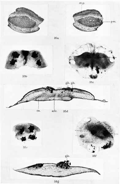
Fig. 35.
a, transverse section through the ventro-lateral body wall of an 8-day budgerigar embryo (stage 2, Part I). The ventral body wall, including the ventral margins of the sternal plates, has been excised and explanted in vitro. (Zenker; haematoxylin and chromatrop. x51.)
b, the ventral body wall removed from the embryo shown in fig. 35a, photographed 2 hr. after explantation. Four patches of gas black have been laid on the surface of the tissue. ( x 22.)
c, same explant photographed after 48 hr. in vitro. Note that the four patches of gas black have moved almost to the mid-line. The dotted line indicates the level of the section shown in fig. 35d. (x 22.)
d, section of the same explant, fixed after 2 days’ growth. Some of the particles of gas black are incorporated in the skin and some in the underlying connective tissue. The edges of the sternal plates are seen as two rods of cartilage connected by a thick sheet of flattened cells. Note that the margin of the explant extends laterally far beyond the cartilaginous rods. (Zenker; haematoxylin and chromatrop. x51.)
e, explant of supra-costal tissue from the same embryo and grown in the same watch-glass as the preceding explant of ventral body wall. Four patches of gas black have been laid on the ectoderm. Photographed shortly after explantation. ( x 25.)
f, same explant after 48 hr. in vitro. The carbon marks have not moved together. The dotted line indicates the position of the section shown in fig. 35g. (x 22.)
g, section of the same explant fixed after 2 days’ cultivation. Note the carbon particles in the ectoderm and subjacent tissue. (Zenker; haematoxylin and chromatrop. x 51.)
|
b.v. blood vessel ¢. coracoid ca. cartilage dc. degenerate cells ect. ectoderm ep.r. epidermal ridge g.b. gas black h. heart hu. humerus 1.f. intercellular fibres k. keel l.b.w. lateral body wall |
my. myoblasts pch. perichondrium p.m. pectoral muscles r. rib sc. scapula s.f.c. sheet of flattened cells st.d. sternal division of rib st.p. sternal plate v.d. vertebral division of rib v.e.k. ventral extension of keel w.b. wing bud |
| Historic Disclaimer - information about historic embryology pages |
|---|
| Pages where the terms "Historic" (textbooks, papers, people, recommendations) appear on this site, and sections within pages where this disclaimer appears, indicate that the content and scientific understanding are specific to the time of publication. This means that while some scientific descriptions are still accurate, the terminology and interpretation of the developmental mechanisms reflect the understanding at the time of original publication and those of the preceding periods, these terms, interpretations and recommendations may not reflect our current scientific understanding. (More? Embryology History | Historic Embryology Papers) |
Reference
Feli HB. The origin and developmental mechanics of the avian sternum. (1939) Phil. Trans. Roy. Soc. Lon. 229 pp.407-463.
Cite this page: Hill, M.A. (2024, April 25) Embryology Feli1939 plate41.jpg. Retrieved from https://embryology.med.unsw.edu.au/embryology/index.php/File:Feli1939_plate41.jpg
- © Dr Mark Hill 2024, UNSW Embryology ISBN: 978 0 7334 2609 4 - UNSW CRICOS Provider Code No. 00098G
File history
Click on a date/time to view the file as it appeared at that time.
| Date/Time | Thumbnail | Dimensions | User | Comment | |
|---|---|---|---|---|---|
| current | 21:57, 13 November 2018 |  | 1,280 × 1,961 (288 KB) | Z8600021 (talk | contribs) | |
| 21:49, 13 November 2018 |  | 2,016 × 2,916 (430 KB) | Z8600021 (talk | contribs) | {{Historic Disclaimer}} ===Reference=== {{Ref-Feli1939}} {{Footer}} |
You cannot overwrite this file.
File usage
The following page uses this file:
