File:Fawcett1930 fig01.jpg

Original file (800 × 669 pixels, file size: 69 KB, MIME type: image/jpeg)
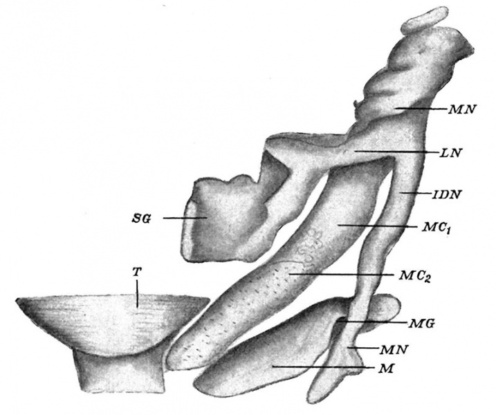
Fig. 1. Reconstruction of left mandible and annexes of a 17 millimetre human embryo viewed from above
Fig. 1 shows the ossified mandible (M) as well as many other structures, e.g. Meckel’s cartilage, which is partially chondrified (M C1) and towards its anterior end unchondrified (M C2); the tongue (T) is also shown near its tip for topographical purposes: finally, the associated nerves, e.g. the mandibular (MN), the lingual (LN) with the submaxillary ganglion (SG), the inferior dental (I DN ), and the mental nerves (MN), are. introduced.
| Historic Disclaimer - information about historic embryology pages |
|---|
| Pages where the terms "Historic" (textbooks, papers, people, recommendations) appear on this site, and sections within pages where this disclaimer appears, indicate that the content and scientific understanding are specific to the time of publication. This means that while some scientific descriptions are still accurate, the terminology and interpretation of the developmental mechanisms reflect the understanding at the time of original publication and those of the preceding periods, these terms, interpretations and recommendations may not reflect our current scientific understanding. (More? Embryology History | Historic Embryology Papers) |
Reference
Fawcett E. A model of the left half of the human mandible at the 17 mm CRL stage. (1930) J Anat Physiol. 47(2): 225-34. PMID:17104287 | PMC1250145
Cite this page: Hill, M.A. (2024, April 19) Embryology Fawcett1930 fig01.jpg. Retrieved from https://embryology.med.unsw.edu.au/embryology/index.php/File:Fawcett1930_fig01.jpg
- © Dr Mark Hill 2024, UNSW Embryology ISBN: 978 0 7334 2609 4 - UNSW CRICOS Provider Code No. 00098G
File history
Click on a date/time to view the file as it appeared at that time.
| Date/Time | Thumbnail | Dimensions | User | Comment | |
|---|---|---|---|---|---|
| current | 14:36, 22 May 2017 |  | 800 × 669 (69 KB) | Z8600021 (talk | contribs) | |
| 14:35, 22 May 2017 |  | 1,315 × 1,080 (175 KB) | Z8600021 (talk | contribs) | {{Ref-Fawcett1930}} {| class="wikitable mw-collapsible mw-collapsed" ! Online Editor || 90px|left This historic 1930 brief note by Fawcett is an early description of the development of the human mandible using a model reco... |
You cannot overwrite this file.
File usage
The following page uses this file:

