File:Fawcett1910 fig02.jpg
From Embryology
Size of this preview: 654 × 600 pixels. Other resolution: 1,035 × 949 pixels.
Original file (1,035 × 949 pixels, file size: 265 KB, MIME type: image/jpeg)
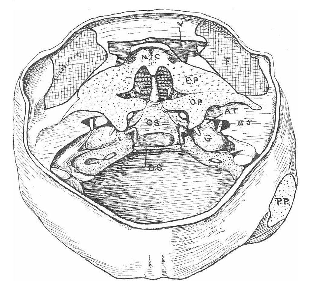
Fig. 2. View from above of the interior of the base of the neuro-cranium of the Bryce 30 mm embryo
The cartilage is everywhere "stippled" and crosses are placed on the ethmoidal plate.
- A.T. - ala temporalis or pars zygomatica of great wing
- C.S. - corpus sphenoidale
- D.S. - dorsum sellae
- E.P. - ethmoidal plate continuous in front with nasal capsule (N.C.) and behind with ala orhitalis (O.P.)
- the fissure at the inner side of the junction of ala orbitalis and ethmoidal plate is the spheno-ethmoidal fissure
- F. - frontal bone
- PP. - parietal plate
- V. - vacuity owing to large blood-vessel or perhaps extravasation.
Links: Fig 1 | Fig 2 | Fig 3 | Fig 4 | Skull Development | Carnegie stage 23
| Historic Disclaimer - information about historic embryology pages |
|---|
 |
- Edward Fawcett Links: 1906 Palate | 1910 Head | 1910 Sphenoid | 1911 Maxilla, vomer, and paraseptal cartilages | 1913 Clavicle | 1930 Mandible | Fawcett image | Edward Fawcett
Reference
Fawcett E. Description of a reconstruction of the head of a thirty-millimetre embryo. (1910) J Anat. Physiol. 44(4): 303-11.
Cite this page: Hill, M.A. (2024, April 20) Embryology Fawcett1910 fig02.jpg. Retrieved from https://embryology.med.unsw.edu.au/embryology/index.php/File:Fawcett1910_fig02.jpg
- © Dr Mark Hill 2024, UNSW Embryology ISBN: 978 0 7334 2609 4 - UNSW CRICOS Provider Code No. 00098G
File history
Click on a date/time to view the file as it appeared at that time.
| Date/Time | Thumbnail | Dimensions | User | Comment | |
|---|---|---|---|---|---|
| current | 06:12, 27 December 2014 | | 1,035 × 949 (265 KB) | Z8600021 (talk | contribs) | |
| 06:10, 27 December 2014 | 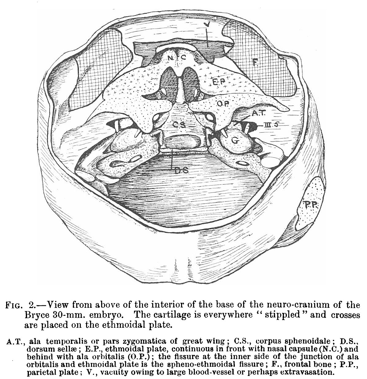 | 1,194 × 1,234 (381 KB) | Z8600021 (talk | contribs) |
You cannot overwrite this file.
File usage
The following page uses this file: