File:Ewart1897 02.jpg
Ewart1897_02.jpg (555 × 600 pixels, file size: 44 KB, MIME type: image/jpeg)
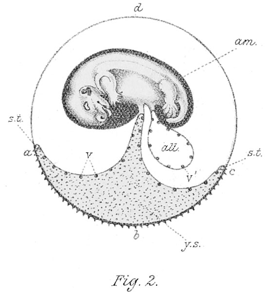
Fig. 2. Represents a young opossum and its foetal appendages
The wall of the yolk sac (y.s.) is vascular as far as the circular blood vessel (s.t., sinus terminalis). In the area a, b, c, the yolk sac blends with the outer embryonic sac. Through this area the cells (trophoblastic) of the outer sac are modified so as to assist in taking up nourishment— the uterine milk — from the uterus, and in fixing the embryo during its uterine development. The allantois (all.) never reaches the outer sac. It is vascular, and serves only as a breathing organ.
After Osborn and Selenka.
| Historic Disclaimer - information about historic embryology pages |
|---|
| Pages where the terms "Historic" (textbooks, papers, people, recommendations) appear on this site, and sections within pages where this disclaimer appears, indicate that the content and scientific understanding are specific to the time of publication. This means that while some scientific descriptions are still accurate, the terminology and interpretation of the developmental mechanisms reflect the understanding at the time of original publication and those of the preceding periods, these terms, interpretations and recommendations may not reflect our current scientific understanding. (More? Embryology History | Historic Embryology Papers) |
Reference
Ewart, J.C. A Critical Period in the Development of the Horse. London: Adam and Charles Black (1897).
File history
Click on a date/time to view the file as it appeared at that time.
| Date/Time | Thumbnail | Dimensions | User | Comment | |
|---|---|---|---|---|---|
| current | 16:13, 3 May 2013 |  | 555 × 600 (44 KB) | Z8600021 (talk | contribs) |
You cannot overwrite this file.
File usage
The following page uses this file:

