File:Evans1909 fig09-12.jpg
Original file (3,089 × 1,606 pixels, file size: 290 KB, MIME type: image/jpeg)
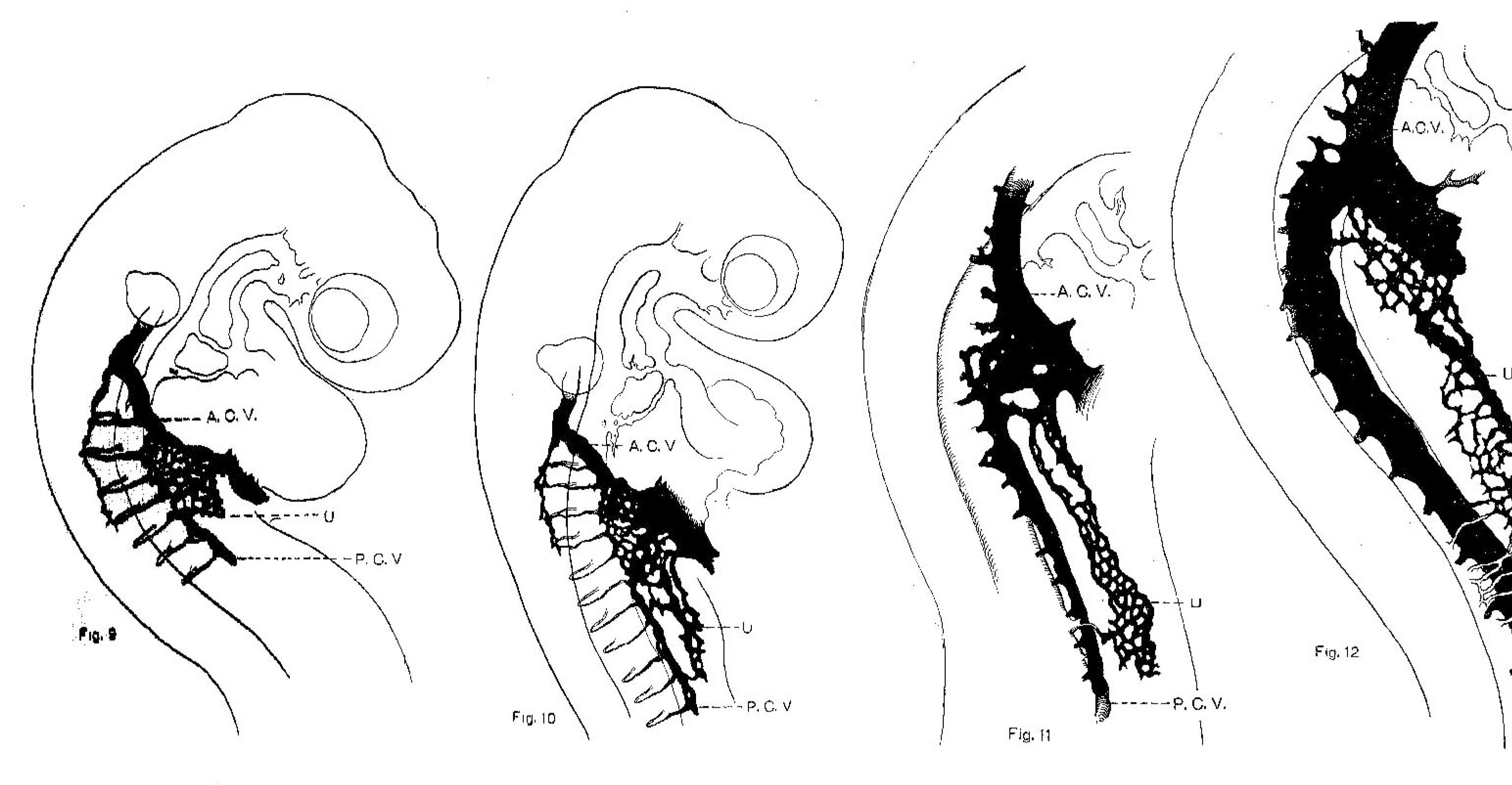
Figs. 9-12.Chick Embryo 23, 24, 30, 35 Somites
Fig. 9. Injected chick embryo of 23 somites to show the origin of the umbilical vein from a capillary plexus situated in the angle between the posterior cardinal vein and the duct of Cuvier. A. C. V. = anterior cardinal vein; P. C. V. = posterlor cardinal vein; U = capiliaries destined to form the umbilical vein.
Fig. 10. Injected chick embryo of 24 somites to show the extension in the somatopleure, of capillary plexus forming the umbilical vein. The lettering is the same as fig. 9.
Fig. 11. Injected chick embryo of 30 somites. The capillaries destined to form the umbilical vein have reached the region of the future arm bud where they are joined by a direct capillary sprout from the aorta (subclavian artery).
Fig. 12. Injected chick embryo of 35 somites, showing establishment of umbilical vein as the main drainage channel of the anterior limb.
| Historic Disclaimer - information about historic embryology pages |
|---|
 |
Evans (1909) Figures: 1 Chick 17 somites | 2 Chick 20 somites | 3 Chick 23 somites | 6 Chick lateral 25 somites |
Reference
Evans HM. On the development of the aortae, cardinal and umbilical veins, and the other blood vessels of vertebrate embryos from capillaries. (1909) Anat. Rec. 3: 498-518.
Cite this page: Hill, M.A. (2024, April 19) Embryology Evans1909 fig09-12.jpg. Retrieved from https://embryology.med.unsw.edu.au/embryology/index.php/File:Evans1909_fig09-12.jpg
- © Dr Mark Hill 2024, UNSW Embryology ISBN: 978 0 7334 2609 4 - UNSW CRICOS Provider Code No. 00098G
File history
Click on a date/time to view the file as it appeared at that time.
| Date/Time | Thumbnail | Dimensions | User | Comment | |
|---|---|---|---|---|---|
| current | 08:02, 29 November 2017 | | 3,089 × 1,606 (290 KB) | Z8600021 (talk | contribs) |
You cannot overwrite this file.