File:Evans1909 fig03b-06.jpg

Original file (3,000 × 2,100 pixels, file size: 457 KB, MIME type: image/jpeg)
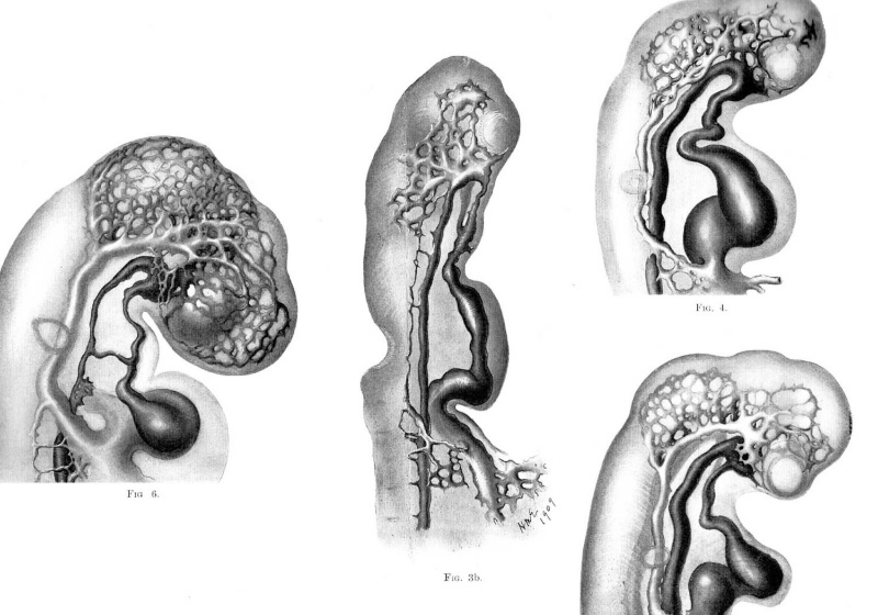
Fig. 3b. Lateral view of head of injected chick embryo of 15 somites, showing primary head capillary plexus. The plexus takes origin from the convexity of the first aortic arch at several points and is continued posteriorly as a slender capillary chain which eventually joins the main vitelline vein near its junction with the heart. This slender capillary chain has arisen at several points from the dorsal aorta on each side, and two of these points of origin are still preserved, opposite the region of the hind brain. The delicate capillary path from the head plexus to the vitelline vein is destined to form the anterior cardinal vein.
Fig. 4. Lateral view of head of injected chick embryo of 17 somites. showing the primary head capillary plexus partially covering the lateral sides of the fore and mid-brain vesicles. It will be seen that a‘ portion of the plexus lies more superficially than the remainder, and it is this superficial portion which is destined to become the main trunk of the vein in this region. The artery is shown darker than the vein.
Fig. 5. Lateral view of head of injected chick embryo of 20 omites. showing the further development of the anterior cardinal vein out of the primary head capillary plexus. The capillaries bordering the groove between mid and bind brain have formed a prominent tributary of the main vein.
Fig. 6. Lateral view of head of injected chick embryo of 25 somites. The lateral surfaces of the fore and mid brain vesicles are now completely covered by the capillary net, which is extending dorsally but is still far from the middorsal line. There is seen a corresponding great growth of the anterior cardinal vein and its system of tributaries.
| Historic Disclaimer - information about historic embryology pages |
|---|
 |
Evans (1909) Figures: 1 Chick 17 somites | 2 Chick 20 somites | 3 Chick 23 somites | 6 Chick lateral 25 somites |
Reference
Evans HM. On the development of the aortae, cardinal and umbilical veins, and the other blood vessels of vertebrate embryos from capillaries. (1909) Anat. Rec. 3: 498-518.
Cite this page: Hill, M.A. (2024, April 24) Embryology Evans1909 fig03b-06.jpg. Retrieved from https://embryology.med.unsw.edu.au/embryology/index.php/File:Evans1909_fig03b-06.jpg
- © Dr Mark Hill 2024, UNSW Embryology ISBN: 978 0 7334 2609 4 - UNSW CRICOS Provider Code No. 00098G
File history
Click on a date/time to view the file as it appeared at that time.
| Date/Time | Thumbnail | Dimensions | User | Comment | |
|---|---|---|---|---|---|
| current | 13:37, 28 November 2017 | | 3,000 × 2,100 (457 KB) | Z8600021 (talk | contribs) |
You cannot overwrite this file.