File:Evans1909 fig02.jpg
Original file (596 × 1,000 pixels, file size: 97 KB, MIME type: image/jpeg)
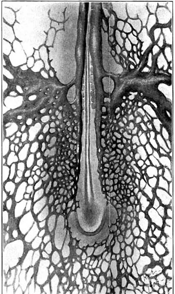
Fig. 2. Ventral view of the posterior part of an injected chick embryo of 20 somites
Showing an extension of the plexus out of which the aorta develops. The large nonvascular area surrounding the region of the primitive steak is here much reduced in extent.
| Historic Disclaimer - information about historic embryology pages |
|---|
 |
Evans (1909) Figures: 1 Chick 17 somites | 2 Chick 20 somites | 3 Chick 23 somites | 6 Chick lateral 25 somites |
Reference
Evans HM. On the development of the aortae, cardinal and umbilical veins, and the other blood vessels of vertebrate embryos from capillaries. (1909) Anat. Rec. 3: 498-518.
Cite this page: Hill, M.A. (2024, April 24) Embryology Evans1909 fig02.jpg. Retrieved from https://embryology.med.unsw.edu.au/embryology/index.php/File:Evans1909_fig02.jpg
- © Dr Mark Hill 2024, UNSW Embryology ISBN: 978 0 7334 2609 4 - UNSW CRICOS Provider Code No. 00098G
File history
Click on a date/time to view the file as it appeared at that time.
| Date/Time | Thumbnail | Dimensions | User | Comment | |
|---|---|---|---|---|---|
| current | 13:27, 28 November 2017 | | 596 × 1,000 (97 KB) | Z8600021 (talk | contribs) |
You cannot overwrite this file.