File:Echidna egg ultrasound.jpg
Echidna_egg_ultrasound.jpg (484 × 390 pixels, file size: 83 KB, MIME type: image/jpeg)
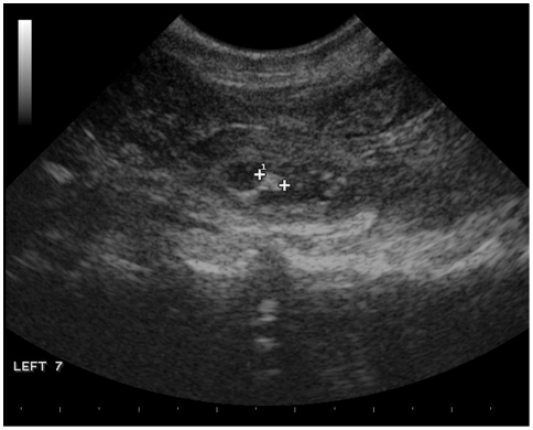
Ultrasound image showing an egg in the uterus of echidna 5D5E on July 23 2008.
Fertilization probably occurred on July 9, but she had fresh sperm in her reproductive tract and was also torpid. Distance between the two markers showing the structure within the egg is 0.35 cm.
Figure 1 http://www.ncbi.nlm.nih.gov/pmc/articles/PMC2699653/figure/pone-0006070-g001/
PLoS One. 2009; 4(6): e6070.
Published online 2009 June 29. doi: 10.1371/journal.pone.0006070.
Copyright Morrow, Nicol. This is an open-access article distributed under the terms of the Creative Commons Attribution License, which permits unrestricted use, distribution, and reproduction in any medium, provided the original author and source are credited.
File history
Click on a date/time to view the file as it appeared at that time.
| Date/Time | Thumbnail | Dimensions | User | Comment | |
|---|---|---|---|---|---|
| current | 09:00, 31 March 2010 |  | 484 × 390 (83 KB) | S8600021 (talk | contribs) | Ultrasound image showing an egg in the uterus of echidna 5D5E on July 23 2008. Fertilization probably occurred on July 9, but she had fresh sperm in her reproductive tract and was also torpid. Distance between the two markers showing the structure within |
You cannot overwrite this file.
File usage
The following page uses this file:
