File:Drosophila stage 1.jpg
From Embryology
No higher resolution available.
Drosophila_stage_1.jpg (425 × 177 pixels, file size: 20 KB, MIME type: image/jpeg)
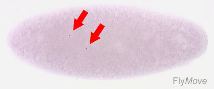
Stage 1 Drosophila Embryo
The two nuclei are marked with arrows following the first cleavage division. A fuchsin stain was used.
Image courtesy of Katrin Weigmann, Robert Klapper, Thomas Strasser, Christof Rickert, Gerd Technau, Herbert Jäckle, Wilfried Janning and Christian Klämbt: FlyMove – a new way to look at development of Drosophila.Trends Genet. In press. http://flymove.uni-muenster.de Image used with permission from Christian Klämbt
- Links: Fly Development
File history
Click on a date/time to view the file as it appeared at that time.
| Date/Time | Thumbnail | Dimensions | User | Comment | |
|---|---|---|---|---|---|
| current | 12:16, 18 September 2009 | | 425 × 177 (20 KB) | Z3217015 (talk | contribs) | The Stage 1 Drosophila Embryo. The two nuclei are marked with arrows following the first cleavage division. A fuchsin stain was used. Image courtesy of Katrin Weigmann, Robert Klapper, Thomas Strasser, Christof Rickert, Gerd Technau, Herbert Jäckle, Wil |
You cannot overwrite this file.
File usage
The following page uses this file: