File:Dog day0-cleft palate.jpg

Original file (490 × 653 pixels, file size: 39 KB, MIME type: image/jpeg)
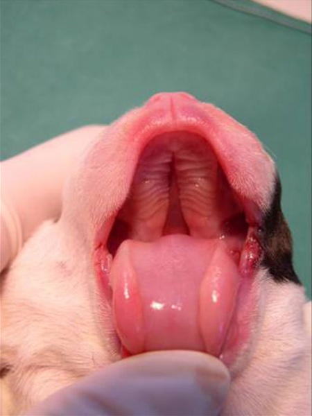
Canine Newborn with Cleft Palate
Bulldog pup (day 0) with cleft palate.
- primary palate (maxilla) has fused
- secondary palate (palatal shelves) have failed to fuse in the midline
- oral cavity abnormally communicates with nasal cavity.
- palatal rugae
- Links: Dog Development | Palate Development
Canine embryo E21 image001
Image Source: Image contributed by Dr Karine Reynaud, Department of Life Sciences and Health and UMR Developmental and Reproduction Biology ENVA, UMR 1198 Biologie du De ́veloppement et Reproduction, 7 Avenue du Ge ́ne ́ral De Gaulle, 94700 Maisons-Alfort, France. CINRA, UMR 1198 Biologie du De ́veloppement et Reproduction, F-78350 Jouy en Josas, France. Who kindly provided these images of the developing dog. Images are for educational purposes only and cannot be reproduced electronically or in writing without permission.
File history
Click on a date/time to view the file as it appeared at that time.
| Date/Time | Thumbnail | Dimensions | User | Comment | |
|---|---|---|---|---|---|
| current | 16:17, 12 April 2011 |  | 490 × 653 (39 KB) | S8600021 (talk | contribs) | ==Canine Newborn (day 0)== Bulldog pup with cleft palate. Canine embryo E21 image001 {{Template:Karine}} Category:Palate |
You cannot overwrite this file.
File usage
The following 5 pages use this file: