File:Dixon1927 fig01-05.jpg

Original file (1,642 × 2,459 pixels, file size: 223 KB, MIME type: image/jpeg)
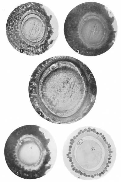
Figs. 1-5
The four photographs, 1, 2, 3 and 4, were taken at different depths of focus. In each the oölemma, the nucleus of the first polar body, the perivitelline space and cytoplasm of the oöcyte are shown, also the cells of the cumulus oöphoros.
Fig. 1 shows the ovoid outline of the oölemma and the more circular form of the cytoplasm as seen in section. The minute irregular intervals and the fine fibrils which cross the space between the oölemma and the cells of the cumulus are seen except on the right side of the photograph. [x300 approx.]
Fig. 2 shows in addition some of the chromosomes of the oöcyte; the narrow cleft where the perivitelline substance has contracted away from the cytoplasm is seen on the right side of the photograph. [x300 approx.]
Fig. 3 under higher magnification than the other photographs. The nucleus and nucleolus of the polar body are seen in more detail. In the space surrounding the oölemma the radiating fibrils, some of which are cut transversely, are seen. [x560 approx.]
Fig. 4. The microscope has been focussed to show some of the individual chromosomes of the dividing nucleus of the oöcyte. x300 approx.
Fig. 5. Semi-diagrammatic sketch made by combining various optical sections. [X400 approx.]
| Historic Disclaimer - information about historic embryology pages |
|---|
| Pages where the terms "Historic" (textbooks, papers, people, recommendations) appear on this site, and sections within pages where this disclaimer appears, indicate that the content and scientific understanding are specific to the time of publication. This means that while some scientific descriptions are still accurate, the terminology and interpretation of the developmental mechanisms reflect the understanding at the time of original publication and those of the preceding periods, these terms, interpretations and recommendations may not reflect our current scientific understanding. (More? Embryology History | Historic Embryology Papers) |
Reference
Dixon AF. Human oocyte showing first polar body and metaphase stage in formation of second polar body. (1927) Irish Jour. Med. Sci., 53: 149-151.
Cite this page: Hill, M.A. (2024, April 25) Embryology Dixon1927 fig01-05.jpg. Retrieved from https://embryology.med.unsw.edu.au/embryology/index.php/File:Dixon1927_fig01-05.jpg
- © Dr Mark Hill 2024, UNSW Embryology ISBN: 978 0 7334 2609 4 - UNSW CRICOS Provider Code No. 00098G
File history
Click on a date/time to view the file as it appeared at that time.
| Date/Time | Thumbnail | Dimensions | User | Comment | |
|---|---|---|---|---|---|
| current | 16:18, 29 November 2017 |  | 1,642 × 2,459 (223 KB) | Z8600021 (talk | contribs) | {{Ref-Dixon1927}} |
You cannot overwrite this file.
File usage
The following page uses this file:
