File:Cytoplasmic lattices in oocytes and two-cell embryos.jpg
Original file (753 × 1,000 pixels, file size: 226 KB, MIME type: image/jpeg)
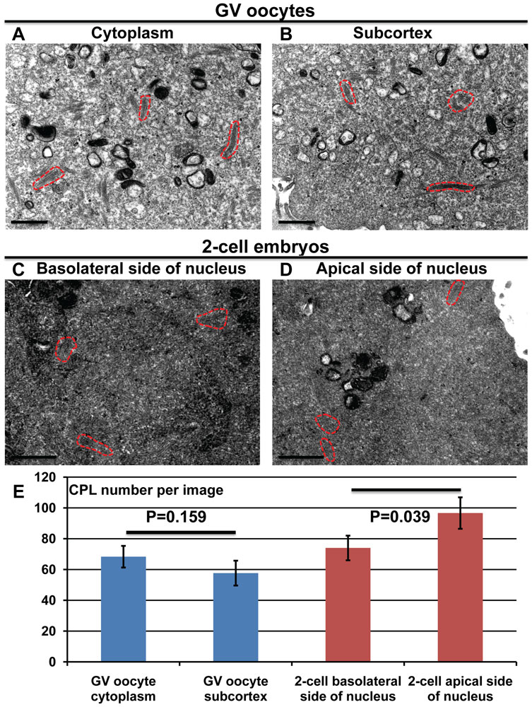
Quantification of cytoplasmic lattices in GV oocytes and two-cell embryos
cytoplasmic lattices (CPLs)
GV oocytes (A and B) and two-cell embryos (C and D) were prepared for TEM. Representative images in the cytoplasm near the nucleus (A) or at the subcortex (B) of GV oocytes, and on either the basolateral (C) or apical (D) side of the nucleus in two-cell embryos. (E) The graph indicates the average number of CPLs per image in the four different aforementioned regions, as well as the two-tailed t-test P values. Representative CPLs are outined in dashed red lines. Bars, 1 µm.
Original file name: Figure 5. Journal.pone.0017226.g005.png
Reference
<pubmed>21359190</pubmed>| PLoS One.
Copyright: © 2011 Morency et al. This is an open-access article distributed under the terms of the Creative Commons Attribution License, which permits unrestricted use, distribution, and reproduction in any medium, provided the original author and source are credited.
File history
Click on a date/time to view the file as it appeared at that time.
| Date/Time | Thumbnail | Dimensions | User | Comment | |
|---|---|---|---|---|---|
| current | 00:01, 24 March 2011 | | 753 × 1,000 (226 KB) | S8600021 (talk | contribs) | ==Quantification of cytoplasmic lattices in GV oocytes and two-cell embryos== cytoplasmic lattices (CPLs) GV oocytes (A and B) and two-cell embryos (C and D) were prepared for TEM. Representative images in the cytoplasm near the nucleus (A) or at the su |
You cannot overwrite this file.
File usage
There are no pages that use this file.