File:Cullen1916 fig93.jpg
Original file (600 × 620 pixels, file size: 73 KB, MIME type: image/jpeg)
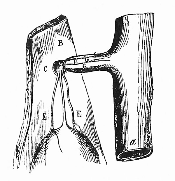
Fig. 93. Meckel's Diverticulum attached to the abdominal wall at the umbilicus
(After Beck.) The picture shows the inner surface of the anterior abdominal wall, to which Meckel's diverticulum has become attached, a is the small bowel; B, the inner surface of the abdominal wall; C, the umbilicus. In the lower part of the picture is seen the bladder. Passing upward from the vertex of this is the urachus. E, E, are the umbilical arteries seen on either side. Passing outward from the small bowel to the umbilicus is Meckel's diverticulum. G, G, represent the omphalomesenteric arteries; H, the omphalomesenteric vein.
| Historic Disclaimer - information about historic embryology pages |
|---|
 |
- Meckel's Diverticulum Links: 92 Meckel's Diverticulum | 93 Meckel's Diverticulum attached to abdominal wall | 94 Large Meckel's Diverticulum | 95 Lobulated Extremity | 96 Hernial Protrusions | 97 Short Meckel's Diverticulum | 98 Accessory Pancreas | 99 Meckel's Diverticulum Tying Loop of Small Bowel | 100 Diverticulum Tying off Loop of Small Bowel | 101 Ileum Volvulus | 102 Fatal Intestinal Obstruction | 103 Meckel's Diverticulum Inversion | Figures | Meckel's diverticulum
Reference
Cullen TS. Embryology, anatomy, and diseases of the umbilicus together with diseases of the urachus. (1916) W. B. Saunders Company, Philadelphia And London.
Cite this page: Hill, M.A. (2024, April 25) Embryology Cullen1916 fig93.jpg. Retrieved from https://embryology.med.unsw.edu.au/embryology/index.php/File:Cullen1916_fig93.jpg
- © Dr Mark Hill 2024, UNSW Embryology ISBN: 978 0 7334 2609 4 - UNSW CRICOS Provider Code No. 00098G
File history
Click on a date/time to view the file as it appeared at that time.
| Date/Time | Thumbnail | Dimensions | User | Comment | |
|---|---|---|---|---|---|
| current | 15:09, 28 October 2018 | | 600 × 620 (73 KB) | Z8600021 (talk | contribs) | |
| 15:07, 28 October 2018 | 664 × 1,694 (225 KB) | Z8600021 (talk | contribs) |
You cannot overwrite this file.
File usage
The following 3 pages use this file: