File:Cullen1916 fig13.jpg
Original file (1,280 × 1,921 pixels, file size: 276 KB, MIME type: image/jpeg)
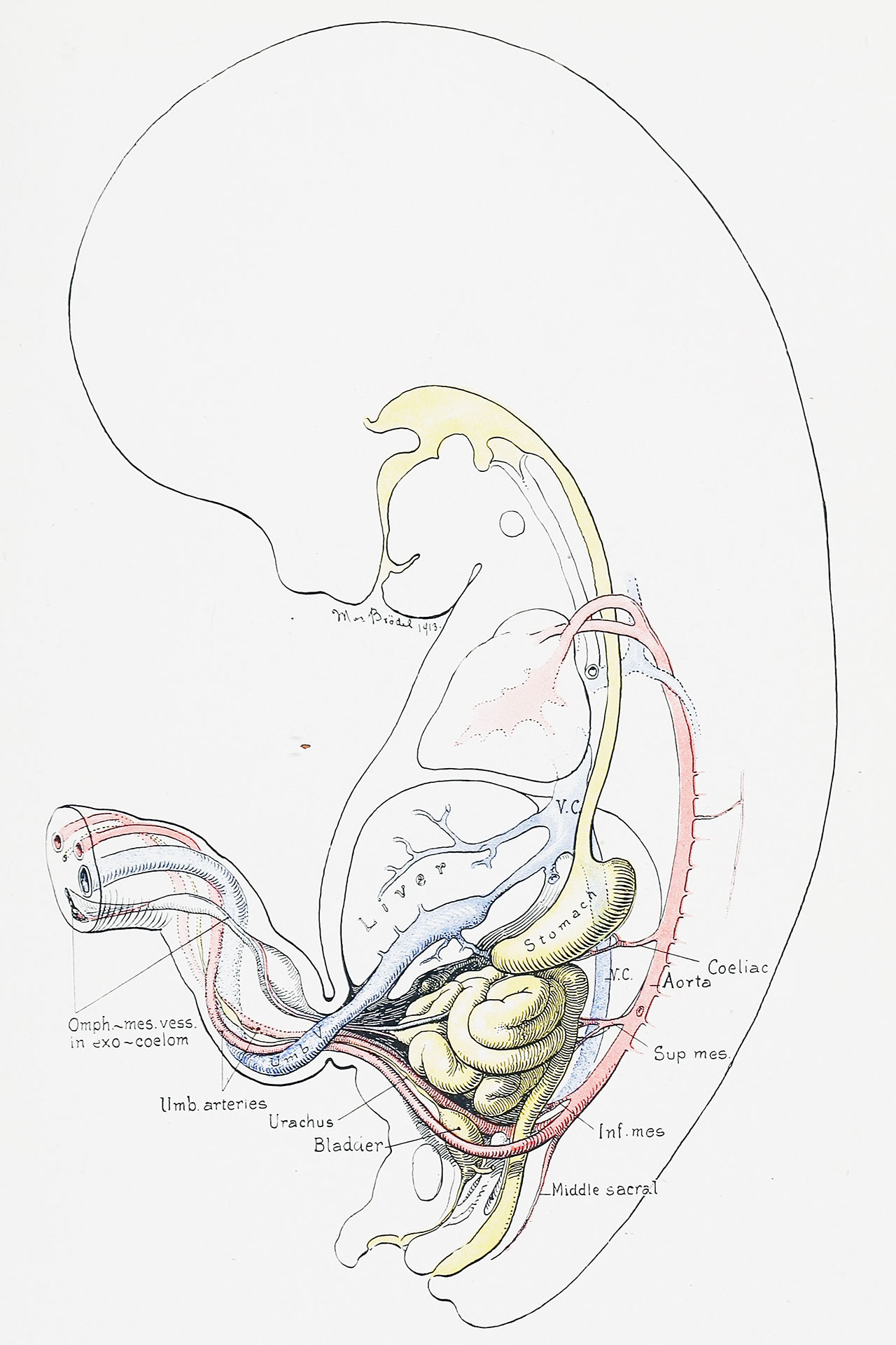
Fig. 13. Sagittal Section of the Umbilical Region in a Human Embryo 4.5 cm in Length
The intestine has receded completely into the abdominal cavity. The exoccelomic cavity, although small in size is still clearly recognizable in the cord, where it shows spindle-shaped dilatations. It now contains only the omphalomesenteric vessels, which have become embedded in its wall. The allantois (urachus) is the distal prolongation of the urinary bladder, and shows already the characteristic dilatations noticeable in older embryos and occasionally found after birth. The structures of the cord show the characteristic twisting of the embryo to the right.
| Historic Disclaimer - information about historic embryology pages |
|---|
 |
- Figure Links: 1 Human embryo 0.7 mm | 2 Human embryo 1.7 mm | 3 Human embryo 2.5 mm | 4 Human embryo 3.5 mm | 5 Human embryo 5 mm | 6 Human embryo 7 mm | 7 Human embryo 7 mm | 8 Human embryo 10 mm | 9 Human embryo 12.5 mm | 10 Human embryo 10 mm | 11 Human embryo 23 mm | 12 Human embryo 3 cm | 13 Human embryo 4.5 cm sagittal | 14 Human Embryo 4.5 cm | 15 Human Embryo 5.2 cm | 16 Human Embryo 6.5 cm | 17 Human Embryo 7.5 cm | 18 Human Embryo 9 cm | 19 Human Embryo 10 cm | 20 Human Embryo 12 cm | 21 Human Embryo 12 cm | 22 Human Embryo 12 cm | 23 Human Embryo 12 cm Cord | 28 Fetus Five Months | 30 Ventral Heria | 31 Human Embryo 5.5 cm | 32 Term Human | 33 Term Human | [[Figures
Reference
Cullen TS. Embryology, anatomy, and diseases of the umbilicus together with diseases of the urachus. (1916) W. B. Saunders Company, Philadelphia And London.
Cite this page: Hill, M.A. (2024, April 24) Embryology Cullen1916 fig13.jpg. Retrieved from https://embryology.med.unsw.edu.au/embryology/index.php/File:Cullen1916_fig13.jpg
- © Dr Mark Hill 2024, UNSW Embryology ISBN: 978 0 7334 2609 4 - UNSW CRICOS Provider Code No. 00098G
File history
Click on a date/time to view the file as it appeared at that time.
| Date/Time | Thumbnail | Dimensions | User | Comment | |
|---|---|---|---|---|---|
| current | 17:25, 27 October 2018 | | 1,280 × 1,921 (276 KB) | Z8600021 (talk | contribs) | |
| 17:24, 27 October 2018 | 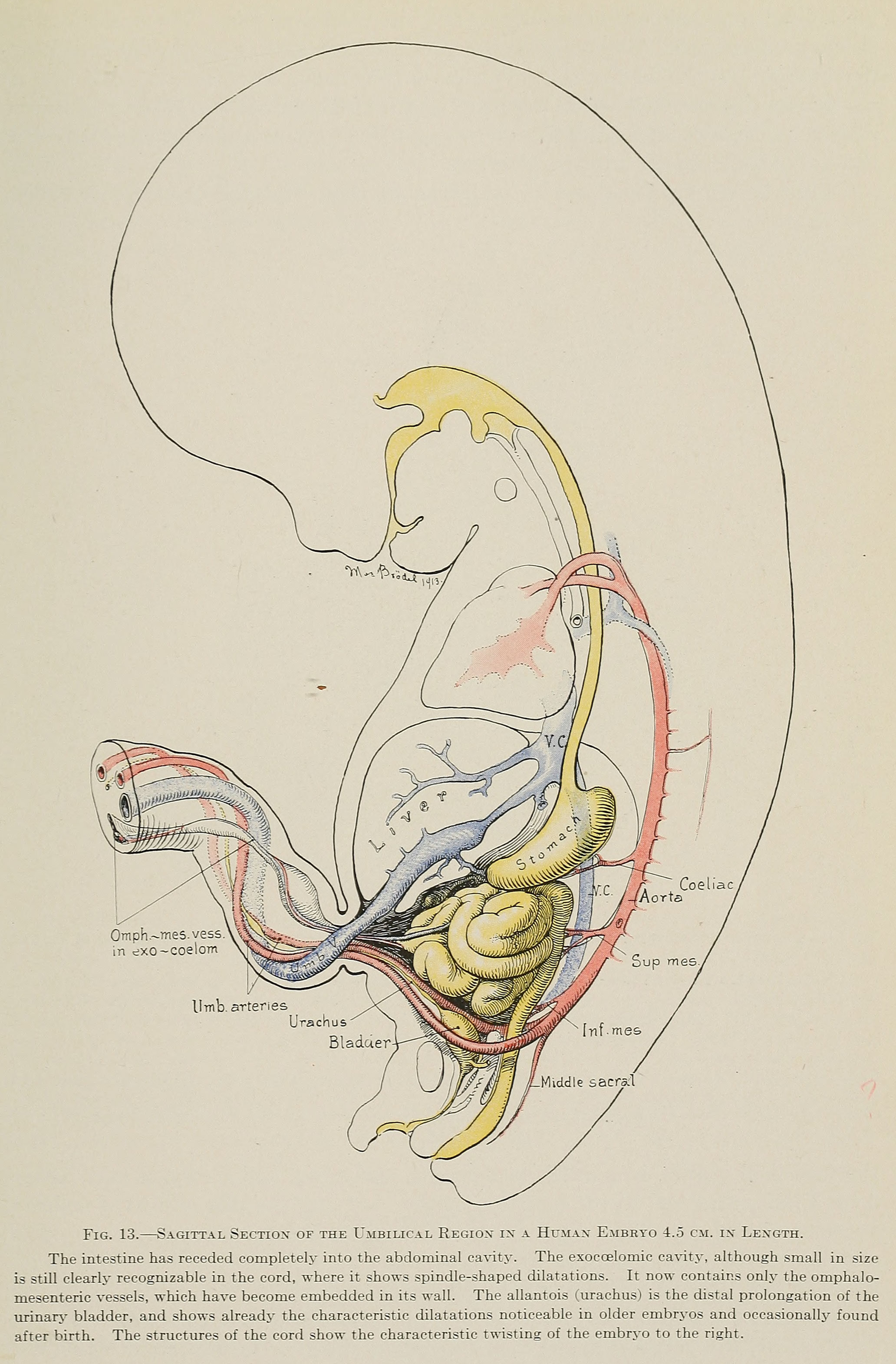 | 2,064 × 3,141 (771 KB) | Z8600021 (talk | contribs) | Fig. 13. Sagittal Section of the Umbilical Region in a Human Embryo 4.5 cm. in Length. The intestine has receded completely into the abdominal cavity. The exoccelomic cavity, although small in size is still clearly recognizable in the cord, where it... |
You cannot overwrite this file.
File usage
The following 3 pages use this file: