File:Cullen1916 fig12.jpg
Original file (1,280 × 1,246 pixels, file size: 560 KB, MIME type: image/jpeg)
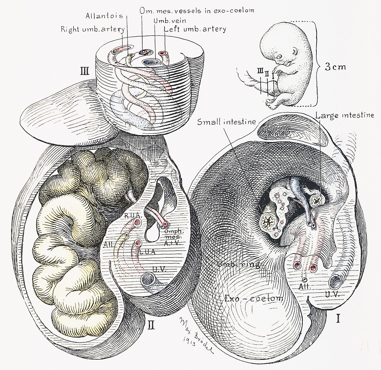
Fig. 12. A Graphic Reconstruction of the Umbilical Region of a Human Embryo 3 cm Long
(Mall collection, No. 86.)
The small sketch in the right upper corner indicates the points at which the sections of the cord have been made. This stage marks the maximal development of the exoccelom, the intestine completely filling its cavity. Section I, the nearest to the embryo, gives a frontal view of the narrow coelomic ring, which, on account of the solid tissue containing i he vascular structures below, appears in the form of a crescent. Emerging from the ring we see the small intestine to the right of the embryo, and the large intestine to the left, with the accompanying mesentery between them. In the cross-section of the mesentery are seen numerous sections of its arteries and vein. The omphalomesenteric vein passes outside of and above the mesentery; the omphalomesenteric artery comes from below, as seen in Section II. They soon leave the intestine and pass out into the funnel-shaped exocoelom.
| Historic Disclaimer - information about historic embryology pages |
|---|
 |
- Figure Links: 1 Human embryo 0.7 mm | 2 Human embryo 1.7 mm | 3 Human embryo 2.5 mm | 4 Human embryo 3.5 mm | 5 Human embryo 5 mm | 6 Human embryo 7 mm | 7 Human embryo 7 mm | 8 Human embryo 10 mm | 9 Human embryo 12.5 mm | 10 Human embryo 10 mm | 11 Human embryo 23 mm | 12 Human embryo 3 cm | 13 Human embryo 4.5 cm sagittal | 14 Human Embryo 4.5 cm | 15 Human Embryo 5.2 cm | 16 Human Embryo 6.5 cm | 17 Human Embryo 7.5 cm | 18 Human Embryo 9 cm | 19 Human Embryo 10 cm | 20 Human Embryo 12 cm | 21 Human Embryo 12 cm | 22 Human Embryo 12 cm | 23 Human Embryo 12 cm Cord | 28 Fetus Five Months | 30 Ventral Heria | 31 Human Embryo 5.5 cm | 32 Term Human | 33 Term Human | [[Figures
Reference
Cullen TS. Embryology, anatomy, and diseases of the umbilicus together with diseases of the urachus. (1916) W. B. Saunders Company, Philadelphia And London.
Cite this page: Hill, M.A. (2024, April 18) Embryology Cullen1916 fig12.jpg. Retrieved from https://embryology.med.unsw.edu.au/embryology/index.php/File:Cullen1916_fig12.jpg
- © Dr Mark Hill 2024, UNSW Embryology ISBN: 978 0 7334 2609 4 - UNSW CRICOS Provider Code No. 00098G
File history
Click on a date/time to view the file as it appeared at that time.
| Date/Time | Thumbnail | Dimensions | User | Comment | |
|---|---|---|---|---|---|
| current | 17:19, 27 October 2018 | | 1,280 × 1,246 (560 KB) | Z8600021 (talk | contribs) | |
| 17:19, 27 October 2018 | 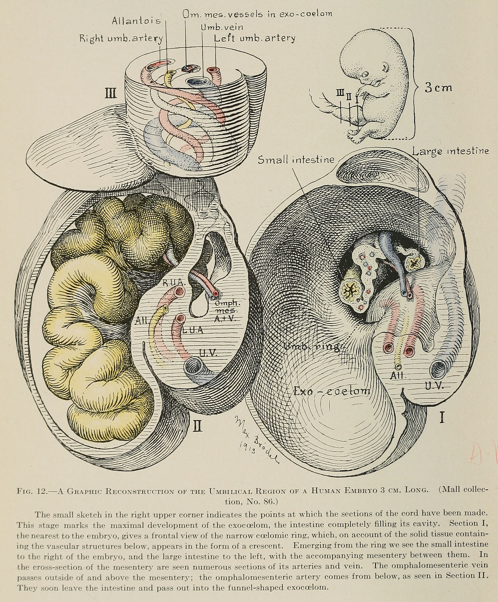 | 2,116 × 2,556 (1.41 MB) | Z8600021 (talk | contribs) | Fig. 12. A Graphic Reconstruction of the Umbilical Region of a Human Embryo 3 cm. Long. (Mall collection, No. 86.) The small sketch in the right upper corner indicates the points at which the sections of the cord have been made. This stage marks the... |
You cannot overwrite this file.
File usage
The following 3 pages use this file: