File:Cullen1916 fig10.jpg
Original file (1,280 × 1,540 pixels, file size: 498 KB, MIME type: image/jpeg)
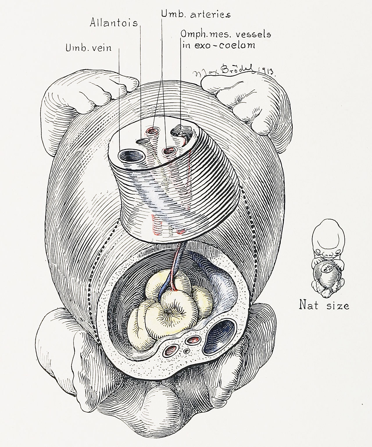
Fig. 10. Anterior View of the Umbilical Cord of a Human Embryo 18 mm in Length
A portion of the cord has been removed, as shown by the dotted line.
The exoccelomic cavity has increased materially in size and contains several loops of intestine, including the cecum and budding appendix, seen on the extreme left (right in picture). The narrow ccelomic ring is obscured by the intestine. The omphalomesenteric duct has disappeared, and we now have only the omphalomesenteric vessels passing out into the exoccelom. This more rapid disappearance of the delicate epithelial structure of the vitelline duct, as contrasted with the preservation of the stronger tissues of the blood-vessels, is probably caused by the continued tension to which the yolk-stalk is subjected. (See Figs. 11 and 12 ) The exocoelomic cavity rapidly tapers to a narrow chink, as seen in the upper and more distal section. In the mid-line of the cord below are the two umbilical arteries, with the allantois between and slightly below them. The umbilical vein lies to the left.
The characteristic twist of the cord, including all of its structures, is clearly seen.
| Historic Disclaimer - information about historic embryology pages |
|---|
 |
- Figure Links: 1 Human embryo 0.7 mm | 2 Human embryo 1.7 mm | 3 Human embryo 2.5 mm | 4 Human embryo 3.5 mm | 5 Human embryo 5 mm | 6 Human embryo 7 mm | 7 Human embryo 7 mm | 8 Human embryo 10 mm | 9 Human embryo 12.5 mm | 10 Human embryo 10 mm | 11 Human embryo 23 mm | 12 Human embryo 3 cm | 13 Human embryo 4.5 cm sagittal | 14 Human Embryo 4.5 cm | 15 Human Embryo 5.2 cm | 16 Human Embryo 6.5 cm | 17 Human Embryo 7.5 cm | 18 Human Embryo 9 cm | 19 Human Embryo 10 cm | 20 Human Embryo 12 cm | 21 Human Embryo 12 cm | 22 Human Embryo 12 cm | 23 Human Embryo 12 cm Cord | 28 Fetus Five Months | 30 Ventral Heria | 31 Human Embryo 5.5 cm | 32 Term Human | 33 Term Human | [[Figures
Reference
Cullen TS. Embryology, anatomy, and diseases of the umbilicus together with diseases of the urachus. (1916) W. B. Saunders Company, Philadelphia And London.
Cite this page: Hill, M.A. (2024, April 23) Embryology Cullen1916 fig10.jpg. Retrieved from https://embryology.med.unsw.edu.au/embryology/index.php/File:Cullen1916_fig10.jpg
- © Dr Mark Hill 2024, UNSW Embryology ISBN: 978 0 7334 2609 4 - UNSW CRICOS Provider Code No. 00098G
File history
Click on a date/time to view the file as it appeared at that time.
| Date/Time | Thumbnail | Dimensions | User | Comment | |
|---|---|---|---|---|---|
| current | 17:12, 27 October 2018 | | 1,280 × 1,540 (498 KB) | Z8600021 (talk | contribs) | |
| 17:11, 27 October 2018 | 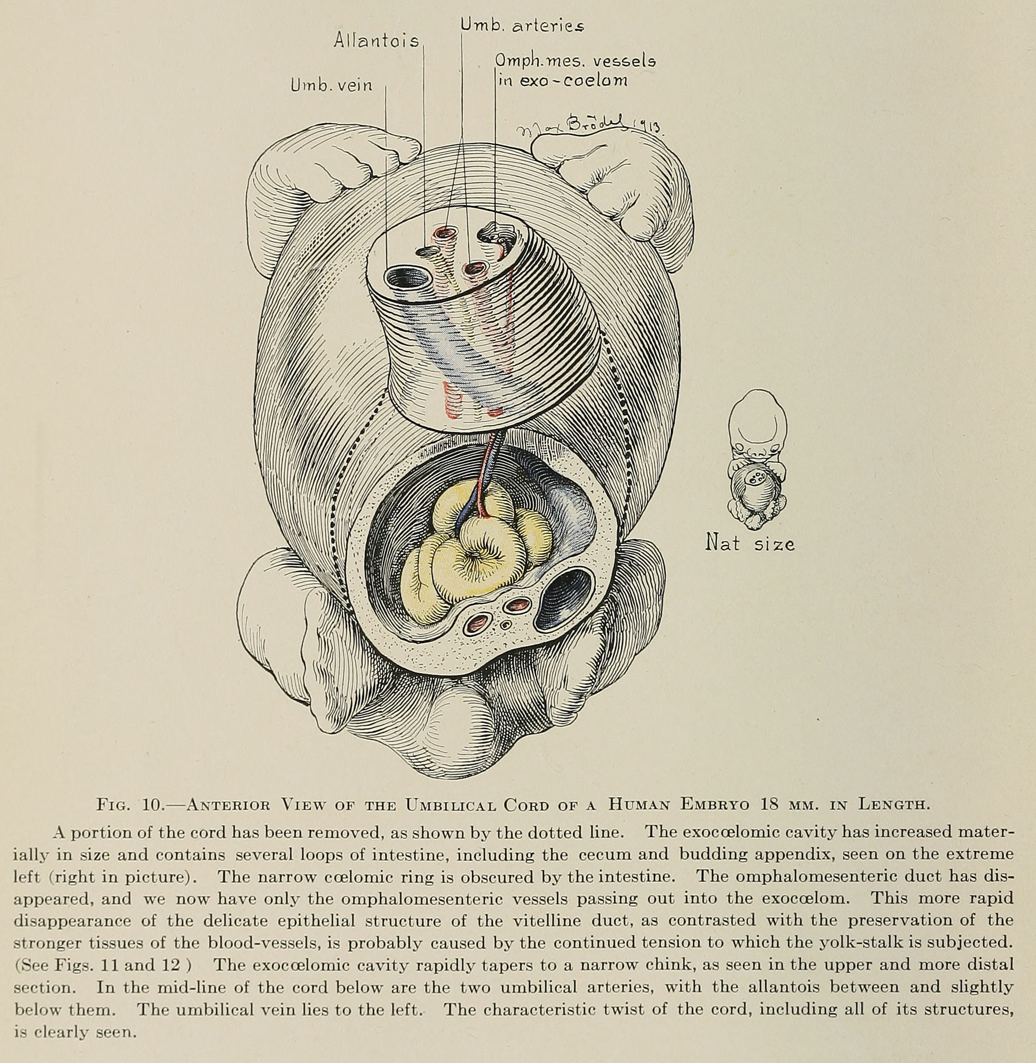 | 2,075 × 2,129 (827 KB) | Z8600021 (talk | contribs) | Fig. 10. Anterior View of the Umbilical Cord of a Human Embryo 18 mm in Length. A portion of the cord has been removed, as shown by the dotted line. The exoccelomic cavity has increased materially in size and contains several loops of intestine, inclu... |
You cannot overwrite this file.
File usage
The following 3 pages use this file: