File:Cullen1916 fig05.jpg
Original file (1,280 × 1,300 pixels, file size: 541 KB, MIME type: image/jpeg)
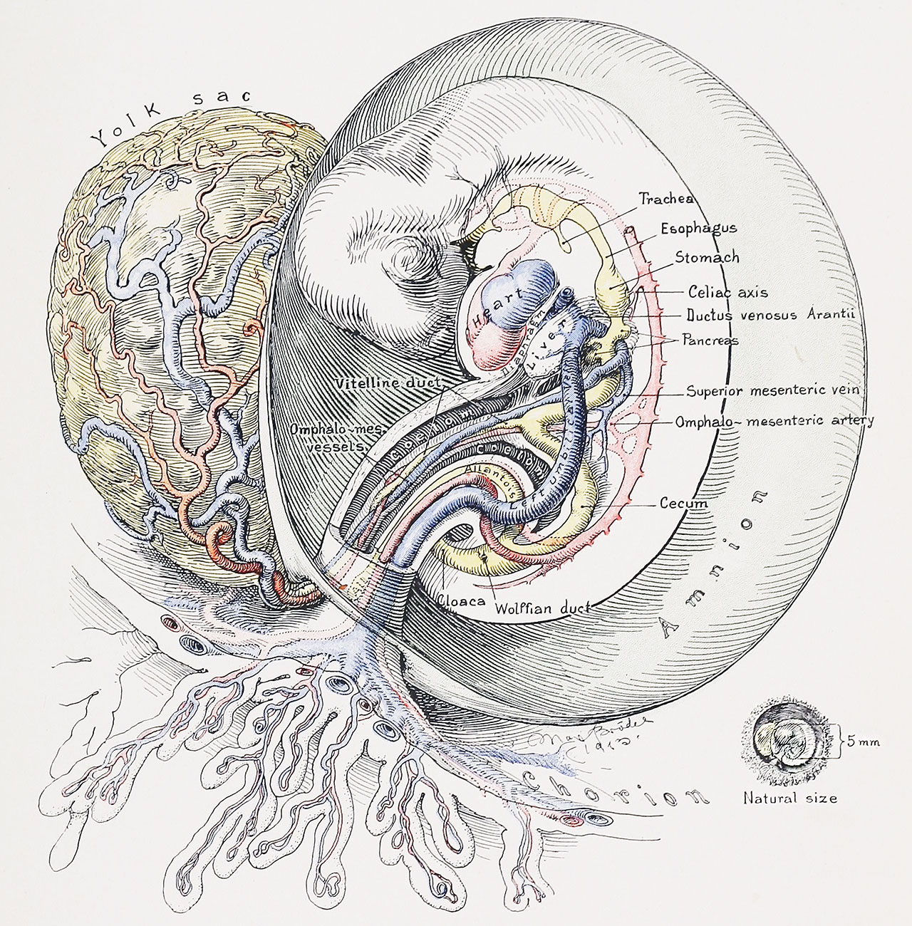
Fig. 5. Sagittal View of a Human Embryo 5 mm in Length
In large part the left halves of the amnion and embryo have been removed, in order to bring clearly into view the structure of the cord. The expanding amnion has almost reached the chorion, hence but little remains of the extra-amniotic portion of the yolk stalk and of the body-stalk. The umbilical cord has become longer. The caudal portion of the cord is firm and contains the umbilical arteries and veins; the latter, soon pfter leaving the body, form a common trunk, which usually curves toward the left. Between the two umbilical arteries lies the allantois, the bulbous end of which still persists almost to the amnion. The cranial portion of the cord is looser in texture. It contains the coelomic cavity, in which lies the omphalomesenteric duct, accompanied by its vessels. Note that the superior mesenteric vein empties into the omphalomesenteric vein behind the pancreatic buds. The omphalomesenteric artery still arises by several branches from the aorta.
| Historic Disclaimer - information about historic embryology pages |
|---|
 |
- Figure Links: 1 Human embryo 0.7 mm | 2 Human embryo 1.7 mm | 3 Human embryo 2.5 mm | 4 Human embryo 3.5 mm | 5 Human embryo 5 mm | 6 Human embryo 7 mm | 7 Human embryo 7 mm | 8 Human embryo 10 mm | 9 Human embryo 12.5 mm | 10 Human embryo 10 mm | 11 Human embryo 23 mm | 12 Human embryo 3 cm | 13 Human embryo 4.5 cm sagittal | 14 Human Embryo 4.5 cm | 15 Human Embryo 5.2 cm | 16 Human Embryo 6.5 cm | 17 Human Embryo 7.5 cm | 18 Human Embryo 9 cm | 19 Human Embryo 10 cm | 20 Human Embryo 12 cm | 21 Human Embryo 12 cm | 22 Human Embryo 12 cm | 23 Human Embryo 12 cm Cord | 28 Fetus Five Months | 30 Ventral Heria | 31 Human Embryo 5.5 cm | 32 Term Human | 33 Term Human | [[Figures
Reference
Cullen TS. Embryology, anatomy, and diseases of the umbilicus together with diseases of the urachus. (1916) W. B. Saunders Company, Philadelphia And London.
Cite this page: Hill, M.A. (2024, April 24) Embryology Cullen1916 fig05.jpg. Retrieved from https://embryology.med.unsw.edu.au/embryology/index.php/File:Cullen1916_fig05.jpg
- © Dr Mark Hill 2024, UNSW Embryology ISBN: 978 0 7334 2609 4 - UNSW CRICOS Provider Code No. 00098G
File history
Click on a date/time to view the file as it appeared at that time.
| Date/Time | Thumbnail | Dimensions | User | Comment | |
|---|---|---|---|---|---|
| current | 16:30, 27 October 2018 | | 1,280 × 1,300 (541 KB) | Z8600021 (talk | contribs) | |
| 16:28, 27 October 2018 | 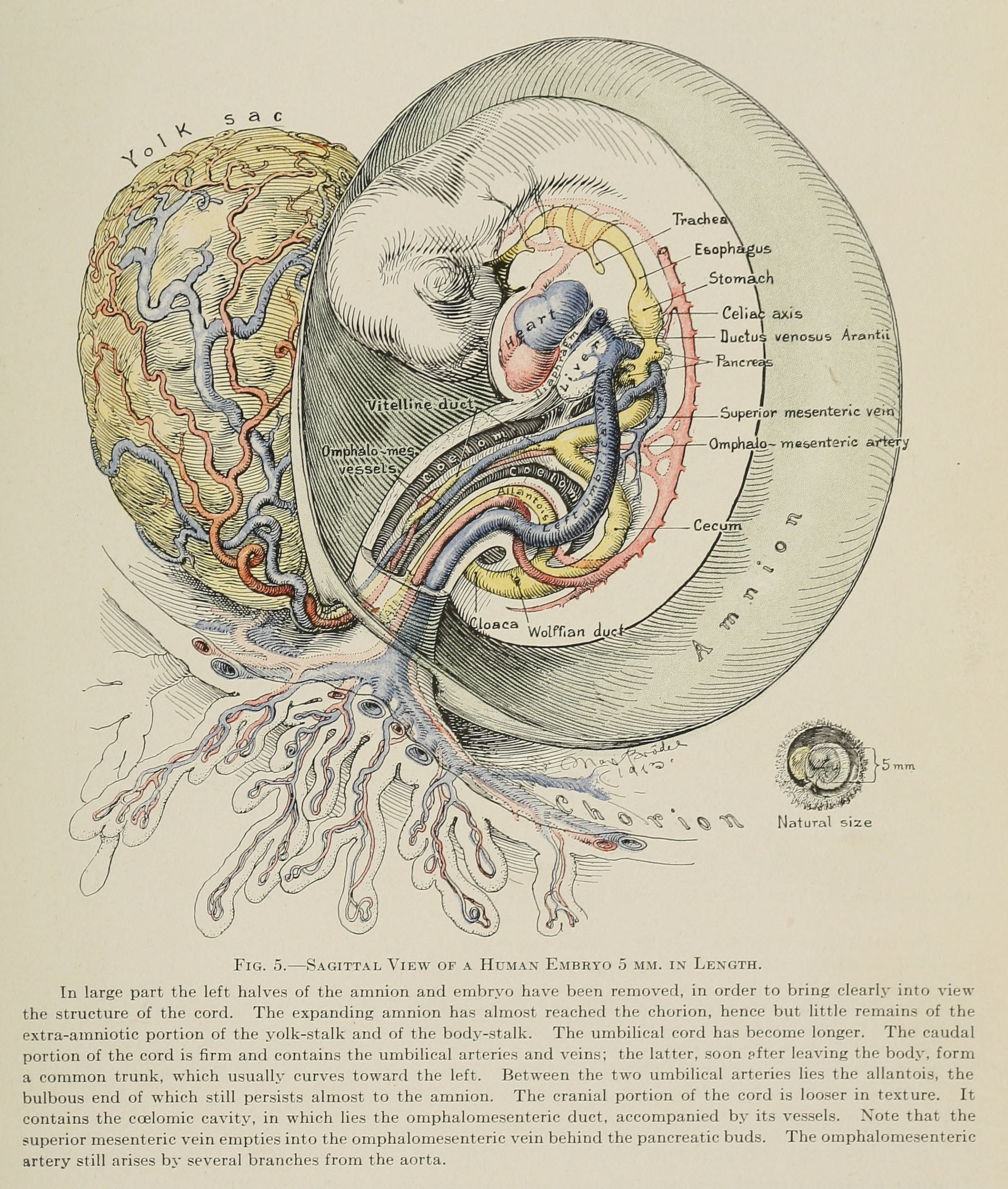 | 2,117 × 2,496 (1.17 MB) | Z8600021 (talk | contribs) |
You cannot overwrite this file.
File usage
The following 3 pages use this file: