File:Conklin 1905 plate09.jpg
Original file (1,521 × 2,000 pixels, file size: 658 KB, MIME type: image/jpeg)
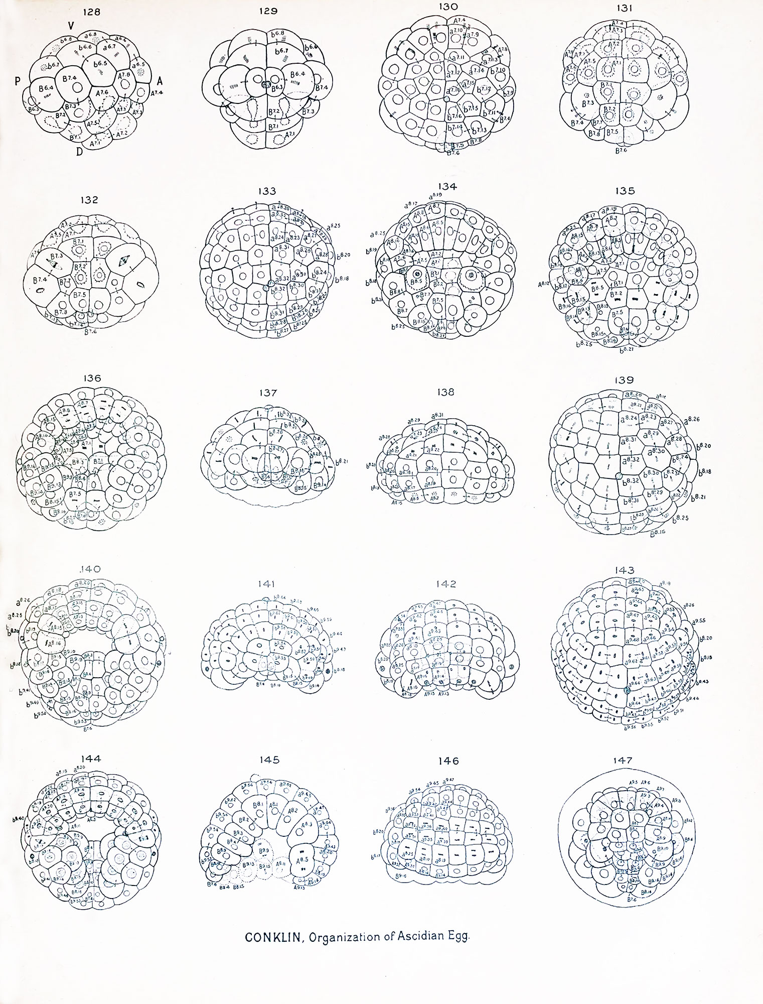
Plate IX Surface Views of Entire Eggs of Cynthia partita - Forty-four to Tiro Hundred and Eighteen Cells
Figs. 128, 129. Same egg as shown in figs. 124-127. Fig. 128 from the left side; the equator of the egg (plane of the third cleavage) is the heavy line running between A ami 1' and separating cells designated by lower case from those designated by capital letters. Fig. 129. View from the posterior pole.
Figs. 130 and 131. Ventral and dorsal views respectively of one and the same egg; 64 cells, 32 in each hemisphere, distributed as follows: Ventral hemisphere, 26 ectoderm, 6 neural plate cells (a 7 -', a 7 -', a 7- ' 3 ) ; Dorsal hemisphere, 10 endoderra, 4 chorda (A 73 , A 77 ), 4 neural plate (A'-, A 7 - 8 ), 10 mesenchyme (B 7 - 6 , B 7 5 , B", B 73 , A 7 ' 6 ) and 4 muscle (B 78 , B 7 ->).
Fig. 132. Postero-dorsal view of an egg in the same stage as the preceding showing direction of division of mesenchyme cell (B 73 ).
Figs. 133 and 134. Ventral and dorsal views respectively of one and the same egg; 110 cells; Ventral hemisphere 64 cells, 52 ectoderm, 12 neural plate (a 8 ' 9 , a 8= , a 8 ' 7 , a 8 ' 8 , a 8,25 , a 8=6 ) ; Dorsal hemisphere 46 cells, 10 endoderm, 8 chorda (A 85 , A 86 , A 8 - 13 , A 8 ' 4 ), 8 neural plate (A 8 - 7 , A 8 - 8 , A 8 -' 3 , A 8 ' 6 ), 12 mesenchyme (B 7 - 6 , B", B 7 7 , B 8 ', B 86 , A 7 - 6 ), 8 muscle (B 8 - 7 , B 88 , B 8 ' 5 , B 8 " 6 ). Gastrulation has begun.
Fig. 135. Dorsal view of a slightly more advanced stage showing increasing gastrulation ; 118 cells, ventral hemisphere 64 cells, dorsal hemisphere 54, viz., 10 endoderm, 16 mesenchyme, 12 muscle, 8 chorda, 8 neural plate; when the divisions indicated by spindles are completed there will 4 additional endoderm and 2 additional mesenchyme cells.
Figs. 136-139. Four views of one and the same egg; fig. 136 dorsal, 137 posterior, 138 anterior, 139 ventral; gastrulation well advanced. 124 cells; Ventral hemisphere 64 cells, 52 ectoderm, 12 neural plate (a 8 -'", a 8 -*", a 8 " 7 , a 8 ' 8 , a 8 - 23 , a 6 - 56 ); Dorsal hemisphere 60 cells, 14 endoderm, 8 chorda, 8 neural plate, 18 mesenchyme, 12 muscle. Spindles are already present for divisions, which, when completed, will lead to 178 cells, viz., 96 ectoderm, 12 neural plate of ventral hemisphere, 12 neural plate of dorsal hemisphere, 8 chorda. 211 mesenchyme, 12 muscle, 18 endoderm.
Figs. 140-143. Four views of one and the same egg; 140 dorsal, 141 posterior, 142 anterior, 143 ventral. Gastrulation is here far advanced. 180 cells; Ventral hemisphere 108 cells, 96 ectoderm, 12 neural plate; Dorsal hemisphere 72 cells, 12 neural plate of dorsal hemisphere, 8 chorda, 20 mesenchyme. 12 muscle, 20 endoderm; when the divisions indicated by spindles are completed there will be 4 additional neural plate cells.
Figs. 144 and 145. Two views of the same egg; 144 dorsal, 145 median optical section in sagittal plane. In fig. 145 the polar body is not visible, but its supposed position is indicated by the dotted outline at the animal pole; the dotted outlines at the lower pole indicate the mesoderm cells which lie in the lateral lip of the blastopore and out of the plane of the section ; the rolling in of the muscle cells in the lateral lips is well shown.
Fig. 146. Anterior view of an egg of about the same stage as the preceding, showing the division of the 12 neural plate cells of the ventral hemisphere.
Fig. 147. Ventral view of a similar stage with the ectoderm omitted in order to show the endoderm and mesoderm from the ventral side. At this stag.' all the ectoderm cells have passed into the 9th generation, all the endoderm into the 8th or 9th, all the mesoderm except IV" into the 8th or 9th, all the chorda and neural plate cells into the 9th. There are 218 cells; Ventral hemisphere 128 cells, 104 ectoderm, 24 neural plate of ventral hemisphere; Dorsal hemisphere 90 cells, viz., 16 mural plate, 16 chorda, 20 mesenchyme, 12 muscle, 26 endoderm.
| Historic Disclaimer - information about historic embryology pages |
|---|
 |
- Conklin Figures: Fig 1-2 | Fig 3-6 | Fig 7-8 | Fig 9-12 | Fig 13-16 | Fig 17-20 | Fig 21-24 | Fig 25-26 | Fig 27-33 | Fig 34-35 | Plate I | Plate II | Plate III | Plate IV | Plate V | Plate VI | Plate VII | Plate VIII | Plate IX | Plate X | Plate XI | Plate XII
Reference
Conklin EG. The Organization and Cell-Lineage of the Ascidian Egg (1905) J. Acad., Nat. Sci. Phila. 13, 1.
Conklin 1905 TOC: I. The Ovarian Egg | II. Maturation and Fertilization | III. Orientation of Egg and Embryo | IV. Cell-Lineage | V. Later Development | VI. Comparisons with A.mphioxus and Amphibia | VII. The Organization of the Egg | Summary | Literature Cited | Explanation of Figures
Cite this page: Hill, M.A. (2024, April 19) Embryology Conklin 1905 plate09.jpg. Retrieved from https://embryology.med.unsw.edu.au/embryology/index.php/File:Conklin_1905_plate09.jpg
- © Dr Mark Hill 2024, UNSW Embryology ISBN: 978 0 7334 2609 4 - UNSW CRICOS Provider Code No. 00098G
File history
Click on a date/time to view the file as it appeared at that time.
| Date/Time | Thumbnail | Dimensions | User | Comment | |
|---|---|---|---|---|---|
| current | 16:26, 19 October 2016 | | 1,521 × 2,000 (658 KB) | Z8600021 (talk | contribs) |
You cannot overwrite this file.
File usage
The following 2 pages use this file: