File:Conklin 1905 plate04.jpg
Original file (1,521 × 2,000 pixels, file size: 429 KB, MIME type: image/jpeg)
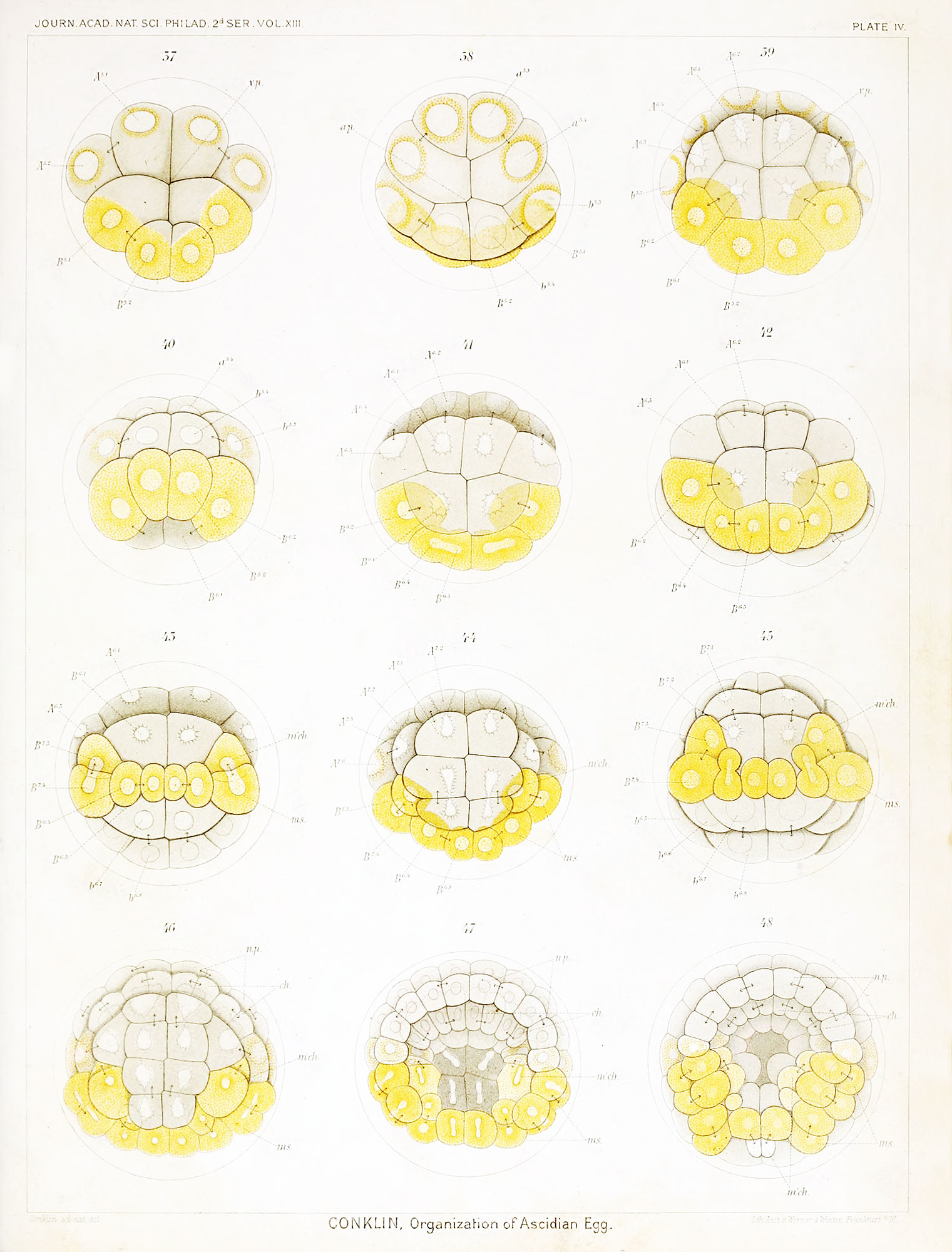
Plate IV Living Eggs of Cynthia partita - Fourth Cleavage to Gastrula
Fig. 37. Sixteen-cell stage viewed from vegetal pole.
Fig. 38. Sixteen-cell stage, from the animal pole, yellow protoplasm around the nuclei.
Fig. 39. Twenty-two cell stage, from the vegetal pole; four mesoderm cells (yellow), ten endoderm, chorda and neural plate cells (gray) and eight ectoderm cells (clear).
Fig. 40. Same stage viewed from the posterior pole.
Fig. 41. Egg passing into the 32-cell stage; postero-dorsal (vegetal pole) view.
Fig. 42. Thirty-two-cell stage, postero-dorsal view.
Fig. 43. Forty-four- cell stage; posterior view, showing separation of mesenchyme (m'ch) from muscle cells (ms.).
Fig. 44. Same stage, dorsal view, showing subdivision of endoderm cells.
Fig. 45. Similar stage, posterior view, showing separation of another mesenchyme cell from a muscle cell.
Fig. 46. Seventy-four cell stage, dorsal view, showing division of 4 chorda and 4 neural piatc cells; there are 10 mesenchyme and 6 muscle cells, besides 10 endoderm cells.
Fig. 47. One hundred and sixteen cell-stage, showing the beginning of gastrulation, also the neural plate, chorda, muscle and mesenchyme cells.
Fig. 48. Slightly older stage showing advancing gastrulation with inrolling of cells at edge of blastopore.
| Historic Disclaimer - information about historic embryology pages |
|---|
 |
- Conklin Figures: Fig 1-2 | Fig 3-6 | Fig 7-8 | Fig 9-12 | Fig 13-16 | Fig 17-20 | Fig 21-24 | Fig 25-26 | Fig 27-33 | Fig 34-35 | Plate I | Plate II | Plate III | Plate IV | Plate V | Plate VI | Plate VII | Plate VIII | Plate IX | Plate X | Plate XI | Plate XII
Reference
Conklin EG. The Organization and Cell-Lineage of the Ascidian Egg (1905) J. Acad., Nat. Sci. Phila. 13, 1.
Conklin 1905 TOC: I. The Ovarian Egg | II. Maturation and Fertilization | III. Orientation of Egg and Embryo | IV. Cell-Lineage | V. Later Development | VI. Comparisons with A.mphioxus and Amphibia | VII. The Organization of the Egg | Summary | Literature Cited | Explanation of Figures
Cite this page: Hill, M.A. (2024, April 19) Embryology Conklin 1905 plate04.jpg. Retrieved from https://embryology.med.unsw.edu.au/embryology/index.php/File:Conklin_1905_plate04.jpg
- © Dr Mark Hill 2024, UNSW Embryology ISBN: 978 0 7334 2609 4 - UNSW CRICOS Provider Code No. 00098G
File history
Click on a date/time to view the file as it appeared at that time.
| Date/Time | Thumbnail | Dimensions | User | Comment | |
|---|---|---|---|---|---|
| current | 15:40, 19 October 2016 | | 1,521 × 2,000 (429 KB) | Z8600021 (talk | contribs) |
You cannot overwrite this file.
File usage
The following 2 pages use this file: