File:Conklin 1905 fig17-20.jpg

Original file (1,000 × 895 pixels, file size: 256 KB, MIME type: image/jpeg)
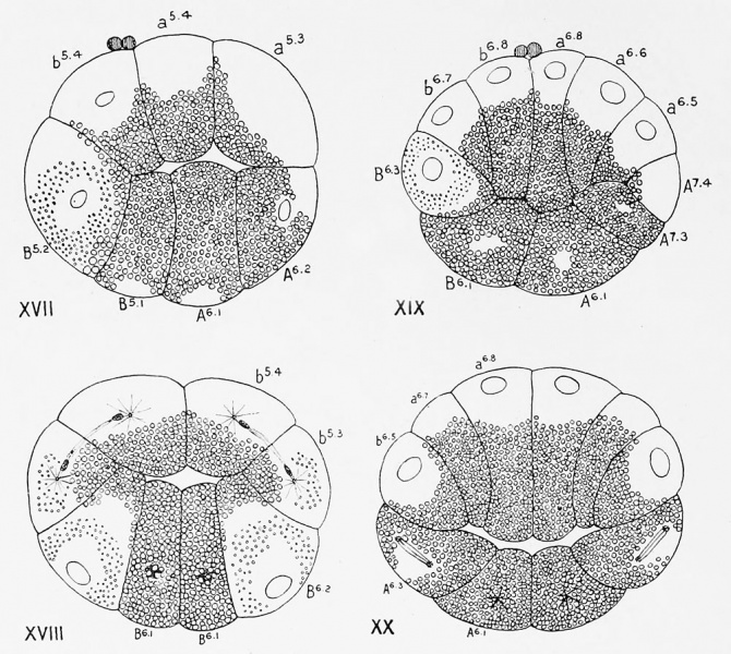
Figs. XVII-XX. Actual sections of eggs of Cynthia partita
Figs. XVII anil XIX in the median plane, Figs. XVIII and XX in a transverse plane. Figs. XVII and XVIII represent a 20-24 cell stage; Figs. XIX and XX a 32-44 cell stage. Unshaded portions of cells represent clear protoplasm ; closely crowded spheres, the yolk ; minute spherules, the yellow protoplasm of the crescent. The clear protoplasm is located chiefly in the cells of the animal half of the egg (ectoderm) ; it is also found in the crescent cells (mesoderm) 1 15; =, B 6! , B 6 '3) and neural plate cells (A7-4, upper half of A 6 -=0 of the lower hemisphere. The remaining cells of the lower hemisphere, (endoderm B5- 1 , B 6 - 1 , A 6,1 , A fc -3, and chorda, A<-3 and lower half of A 6-2 ) are filled with yolk ; yolk is also found in the central ends of all the other cells. The yellow protoplasm is limited to the crescent cells and to a single pair of cells of the upper hemisphere (bs-3). In
Fig. XVII the cells at the two poles are approximately equal in height ; in Fig. XVIII the cells at the
animal pole are flatter, probably owing to the fact that they are dividing ; in Figs. XIX and XX the cells
at the animal pole are columnar, those at the vegetal pole flattened. The polar bodies are actually present where they are represented.
| Historic Disclaimer - information about historic embryology pages |
|---|
| Pages where the terms "Historic" (textbooks, papers, people, recommendations) appear on this site, and sections within pages where this disclaimer appears, indicate that the content and scientific understanding are specific to the time of publication. This means that while some scientific descriptions are still accurate, the terminology and interpretation of the developmental mechanisms reflect the understanding at the time of original publication and those of the preceding periods, these terms, interpretations and recommendations may not reflect our current scientific understanding. (More? Embryology History | Historic Embryology Papers) |
- Conklin Figures: Fig 1-2 | Fig 3-6 | Fig 7-8 | Fig 9-12 | Fig 13-16 | Fig 17-20 | Fig 21-24 | Fig 25-26 | Fig 27-33 | Fig 34-35 | Plate I | Plate II | Plate III | Plate IV | Plate V | Plate VI | Plate VII | Plate VIII | Plate IX | Plate X | Plate XI | Plate XII
Reference
Conklin EG. The Organization and Cell-Lineage of the Ascidian Egg (1905) J. Acad., Nat. Sci. Phila. 13, 1.
Conklin 1905 TOC: I. The Ovarian Egg | II. Maturation and Fertilization | III. Orientation of Egg and Embryo | IV. Cell-Lineage | V. Later Development | VI. Comparisons with A.mphioxus and Amphibia | VII. The Organization of the Egg | Summary | Literature Cited | Explanation of Figures
Cite this page: Hill, M.A. (2024, April 19) Embryology Conklin 1905 fig17-20.jpg. Retrieved from https://embryology.med.unsw.edu.au/embryology/index.php/File:Conklin_1905_fig17-20.jpg
- © Dr Mark Hill 2024, UNSW Embryology ISBN: 978 0 7334 2609 4 - UNSW CRICOS Provider Code No. 00098G
File history
Click on a date/time to view the file as it appeared at that time.
| Date/Time | Thumbnail | Dimensions | User | Comment | |
|---|---|---|---|---|---|
| current | 15:37, 21 October 2016 |  | 1,000 × 895 (256 KB) | Z8600021 (talk | contribs) | |
| 15:37, 21 October 2016 |  | 1,000 × 1,175 (369 KB) | Z8600021 (talk | contribs) |
You cannot overwrite this file.
File usage
The following 2 pages use this file:
