File:Conklin 1905 fig01-02.jpg
Original file (1,000 × 468 pixels, file size: 94 KB, MIME type: image/jpeg)
Fig. 1 - 2
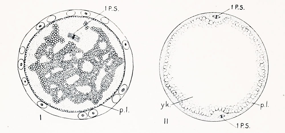
Fig. I. Section of an egg of Cynthia partita which had lain twelve hours without being fertilized. The first polar spindle (1 p. -. lies in tin- position in which it was first formed ; the peripheral layer of yellow protoplasm ip. l.i remains uniformly distributed over the surface, hut the clear protoplasm has spread throughout the yolk and broken it up into irregular masses (compare with figs. 77 and 78 showing unfertilized est:s in normal condition).
Fig. II. Stained preparation of an entire egg of Cynthia partita, showing small spindles at opposite poles (1. p. s.i. which are possibly two first maturation spindles, though more probably one of these is a precociously developed sperm spindle.
| Historic Disclaimer - information about historic embryology pages |
|---|
 |
- Conklin Figures: Fig 1-2 | Fig 3-6 | Fig 7-8 | Fig 9-12 | Fig 13-16 | Fig 17-20 | Fig 21-24 | Fig 25-26 | Fig 27-33 | Fig 34-35 | Plate I | Plate II | Plate III | Plate IV | Plate V | Plate VI | Plate VII | Plate VIII | Plate IX | Plate X | Plate XI | Plate XII
Reference
Conklin EG. The Organization and Cell-Lineage of the Ascidian Egg (1905) J. Acad., Nat. Sci. Phila. 13, 1.
Conklin 1905 TOC: I. The Ovarian Egg | II. Maturation and Fertilization | III. Orientation of Egg and Embryo | IV. Cell-Lineage | V. Later Development | VI. Comparisons with A.mphioxus and Amphibia | VII. The Organization of the Egg | Summary | Literature Cited | Explanation of Figures
Cite this page: Hill, M.A. (2024, April 18) Embryology Conklin 1905 fig01-02.jpg. Retrieved from https://embryology.med.unsw.edu.au/embryology/index.php/File:Conklin_1905_fig01-02.jpg
- © Dr Mark Hill 2024, UNSW Embryology ISBN: 978 0 7334 2609 4 - UNSW CRICOS Provider Code No. 00098G
File history
Click on a date/time to view the file as it appeared at that time.
| Date/Time | Thumbnail | Dimensions | User | Comment | |
|---|---|---|---|---|---|
| current | 16:59, 19 October 2016 | | 1,000 × 468 (94 KB) | Z8600021 (talk | contribs) | |
| 16:59, 19 October 2016 |  | 1,000 × 648 (146 KB) | Z8600021 (talk | contribs) | {{Historic Disclaimer}} {{Conklin1905 figures}} ===Reference=== {{Ref-Conklin1905}} {{Conklin1905 TOC}} {{Footer}} |
You cannot overwrite this file.
File usage
The following 2 pages use this file: