File:Congenital hydrocephalus MRI02.jpg
Congenital_hydrocephalus_MRI02.jpg (595 × 600 pixels, file size: 42 KB, MIME type: image/jpeg)
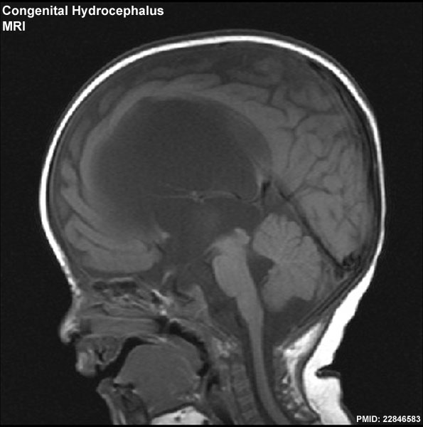
Congenital hydrocephalus (MRI)
Sagittal T1-weighted magnetic resonance image four-month-old male baby demonstrating obstructive hydrocephalus due to congenital aqueductal stenosis.
Dark central region of scan shows the enlarged ventricular region.
- Links: image 1 | image 2 | hydrocephalus
Reference
Bokhari R & Baeesa S. (2012). Remote cerebellar hemorrhage due to ventriculoperitoneal shunt in an infant: a case report. J Med Case Rep , 6, 222. PMID: 22846583 DOI.
Bokhari and Baeesa Journal of Medical Case Reports 2012 6:222 doi:10.1186/1752-1947-6-222
Copyright
© 2012 Bokhari and Baeesa; licensee BioMed Central Ltd. This is an Open Access article distributed under the terms of the Creative Commons Attribution License (http://creativecommons.org/licenses/by/2.0), which permits unrestricted use, distribution, and reproduction in any medium, provided the original work is properly cited.
Original image: Figure 1 (original image size adjusted)
Cite this page: Hill, M.A. (2024, April 25) Embryology Congenital hydrocephalus MRI02.jpg. Retrieved from https://embryology.med.unsw.edu.au/embryology/index.php/File:Congenital_hydrocephalus_MRI02.jpg
- © Dr Mark Hill 2024, UNSW Embryology ISBN: 978 0 7334 2609 4 - UNSW CRICOS Provider Code No. 00098G
File history
Click on a date/time to view the file as it appeared at that time.
| Date/Time | Thumbnail | Dimensions | User | Comment | |
|---|---|---|---|---|---|
| current | 13:02, 7 December 2012 | | 595 × 600 (42 KB) | Z8600021 (talk | contribs) | ==Congenital hydrocephalus (MRI)== Sagittal T1-weighted magnetic resonance image four-month-old male baby demonstrating obstructive hydrocephalus due to congenital aqueductal stenosis. Dark central region of scan shows the enlarged ventricular region. |
You cannot overwrite this file.
File usage
The following 2 pages use this file: