File:Chicken neural tube thickening.jpg
Chicken_neural_tube_thickening.jpg (529 × 600 pixels, file size: 69 KB, MIME type: image/jpeg)
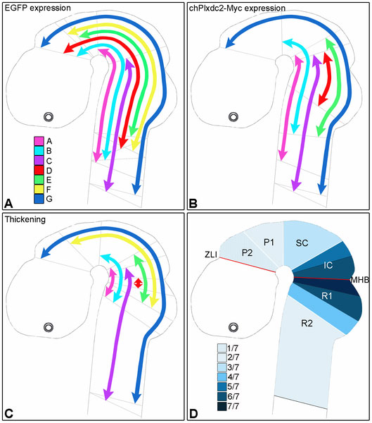
Summary of results from optical projection tomography analysis of chPlxdc2-Myc expression and neural tube thickening
in seven embryos (A–G), 24 hours post electroporation.
OPT - optical projection tomography
a, Extent of EGFP expressing cells;
b, corresponding extent of chPlxdc2-Myc expression (chPlxdc2-Myc data was not obtained for specimen F);
c, extent of neural tube thickening in each of seven specimens;
d, occurence of thickening in individual brain regions.
Grey lines in a-c represent the limits of expression/thickening. IC, inferior colliculus; MHB, midbrain-hindbrain boundary; P1, prosomere 1; P2, prosomere 2; R1, rhombomere 1; R2, rhombomere 2; SC, superior colliculus; ZLI, Zona Limitans Intrathalamica.
Original file name: Figure 5. Journal.pone.0014565.g005.png
Reference
<pubmed>21283688</pubmed>| PMC3024984 | PLoS One
Citation: Miller-Delaney SFC, Lieberam I, Murphy P, Mitchell KJ (2011) Plxdc2 Is a Mitogen for Neural Progenitors. PLoS ONE 6(1): e14565. doi:10.1371/journal.pone.0014565
Copyright: © 2011 Miller-Delaney et al. This is an open-access article distributed under the terms of the Creative Commons Attribution License, which permits unrestricted use, distribution, and reproduction in any medium, provided the original author and source are credited.
File history
Click on a date/time to view the file as it appeared at that time.
| Date/Time | Thumbnail | Dimensions | User | Comment | |
|---|---|---|---|---|---|
| current | 10:49, 20 February 2011 |  | 529 × 600 (69 KB) | S8600021 (talk | contribs) | ==Summary of results from optical projection tomography analysis of chPlxdc2-Myc expression and neural tube thickening== in seven embryos (A–G), 24 hours post electroporation. OPT - optical projection tomography a, Extent of EGFP expressing cells; |
You cannot overwrite this file.
File usage
There are no pages that use this file.
