File:Cervicovaginal mucus rheology.jpg
Cervicovaginal_mucus_rheology.jpg (800 × 438 pixels, file size: 74 KB, MIME type: image/jpeg)
Cervicovaginal Mucus Rheology
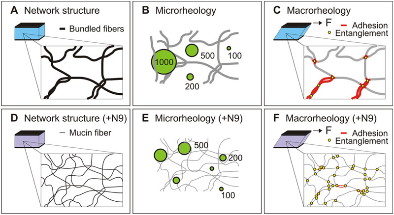
(A) The mesh structure of native human cervicovaginal mucus (CVM) consists of individual mucin fibers bundled together, leading to large mesh spacings.
(B) The microrheology of CVM, quantified using different sized probe nanoparticles, suggests that CVM is largely a viscoelastic fluid at length scales 500 nm or below, whereas at length scales 1,000 nm or higher the mesh elements contribute to a markedly greater local elasticity characteristic of viscoelastic solids.
(C) The macrorheology of CVM reflects contributions from entanglements as well as hydrophobic adhesive interactions between the mesh elements.
(D) Treatment of CVM with nonoxynol-9 (N9) leads to unbundling of the mesh elements and significantly reduced mesh spacings, due to reduced hydrophobic interactions between mucin fibers.
(E) The microrheology of N9-treated CVM becomes that of a viscoelastic solid at length scales down to 200 nm, but remains largely unperturbed at length scales ~100 nm or below.
(F) The effect of N9 cannot be probed by macrorheology, as the reduction in adhesive interactions by N9 is likely balanced by increased entanglements between mucin fibbers.
- "The ability of mucus to function as a protective barrier at mucosal surfaces rests on its viscous and elastic properties, which are not well understood at length scales relevant to pathogens and ultrafine environmental particles. Here we report that fresh, undiluted human cervicovaginal mucus (CVM) transitions from an impermeable elastic barrier to non-adhesive objects sized 1 µm and larger to a highly permeable viscoelastic liquid to non-adhesive objects smaller than 500 nm in diameter. Addition of a nonionic detergent, present in vaginal gels, lubricants and condoms, caused CVM to behave as an impermeable elastic barrier to 200 and 500 nm particles, suggesting that the dissociation of hydrophobically-bundled mucin fibers created a finer elastic mucin mesh. Surprisingly, the macroscopic viscoelasticity, which is critical to proper mucus function, was unchanged. These findings provide important insight into the nanoscale structural and barrier properties of mucus, and how the penetration of foreign particles across mucus might be inhibited."
Original file name: Journal.pone.0004294.g004.png
Reference
Citation: Lai SK, Wang Y-Y, Cone R, Wirtz D, Hanes J (2009) Altering Mucus Rheology to “Solidify” Human Mucus at the Nanoscale. PLoS ONE 4(1): e4294. doi:10.1371/journal.pone.0004294
© 2009 Lai et al. This is an open-access article distributed under the terms of the Creative Commons Attribution License, which permits unrestricted use, distribution, and reproduction in any medium, provided the original author and source are credited.
http://www.plosone.org/article/info%3Adoi%2F10.1371%2Fjournal.pone.0004294
File history
Click on a date/time to view the file as it appeared at that time.
| Date/Time | Thumbnail | Dimensions | User | Comment | |
|---|---|---|---|---|---|
| current | 11:36, 30 July 2011 |  | 800 × 438 (74 KB) | S8600021 (talk | contribs) | ==Cervicovaginal Mucus Rheology== (A) The mesh structure of native human cervicovaginal mucus (CVM) consists of individual mucin fibers bundled together, leading to large mesh spacings. (B) The microrheology of CVM, quantified using different sized prob |
You cannot overwrite this file.
File usage
There are no pages that use this file.
