Cerebral Blood Supply Development
|
Embryonic stage
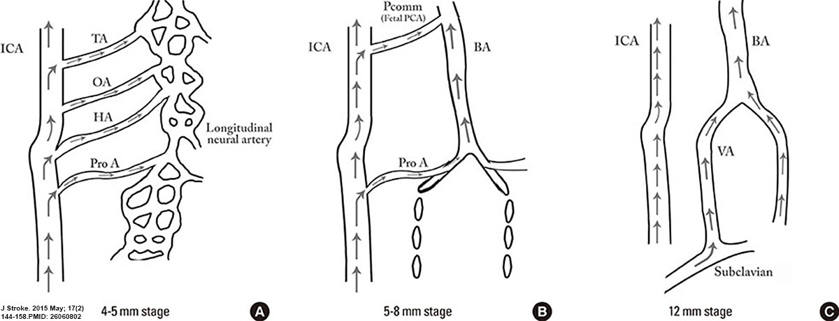
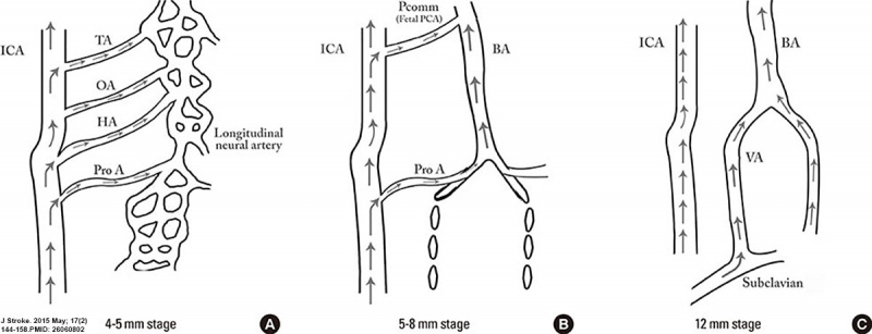
- 4-5 mm - hindbrain (i.e., future posterior fossa) is supplied by two parallel neural arteries (or channels). These arteries obtain their blood supply from carotid-vertebrobasilar anastomoses given by the trigeminal artery (TA), the otic artery (OA), hypoglossal artery (HA), and the proatlantal artery (ProA)
- 5-8 mm - basilar artery (BA) forms from the consolidation of the neural arteries.
- 7-12 mm - vertebral arteries (VA) forms from transverse anastomoses between cervical intersegmental arteries, beginning with the ProA and proceeding downward to the 6th intersegmental artery,
- 11-12 mm - (35 days) development of the MCA is first identified as small buds originating proximal to the ACA on the anterior division of the primitive ICA.
- 16-18 mm - MCA becomes more prominent, the plexi fuse into a single artery and further branches pierce the cerebral hemisphere.
- 18 mm - stem of the ACA gives rise to the olfactory artery.
- 21-24 mm - formation of the anterior communicating artery (ACOMM).
|
- A - In early phases of development the posterior circulation relies almost entirely from blood supply coming from the anterior circulation through carotid-vertebrobasilar anastomoses.
- B and C - As the posterior fossa structures and the occipital lobe grow, the posterior circulation becomes progressively independent from the anterior circulation with obliteration of the anterior-posterior anastomoses from caudal to rostral maintaining in the majority of adult only one connection between the distal basilar arteries with the carotid artery via the posterior communicating artery.
|
(above text modified from reference)
- Links: Neural - Vascular Development | Neural System Development | Cardiovascular System Development | Head Development
Reference
Menshawi K, Mohr JP & Gutierrez J. (2015). A Functional Perspective on the Embryology and Anatomy of the Cerebral Blood Supply. J Stroke , 17, 144-58. PMID: 26060802 DOI.
Copyright
This is an Open Access article distributed under the terms of the Creative Commons Attribution Non-Commercial License (http://creativecommons.org/licenses/by-nc/3.0/) which permits unrestricted non-commercial use, distribution, and reproduction in any medium, provided the original work is properly cited.
Figure 2. http://synapse.koreamed.org/ViewImage.php?Type=F&aid=497461&id=F2&afn=1183_JOS_17_2_144&fn=jos-17-144-g002_1183JOS
Jos-17-144-g002-l.jpg
Cite this page: Hill, M.A. (2024, April 16) Embryology Cerebral blood supply development 01.jpg. Retrieved from https://embryology.med.unsw.edu.au/embryology/index.php/File:Cerebral_blood_supply_development_01.jpg
- What Links Here?
- © Dr Mark Hill 2024, UNSW Embryology ISBN: 978 0 7334 2609 4 - UNSW CRICOS Provider Code No. 00098G
Click on a date/time to view the file as it appeared at that time.
| Date/Time | Thumbnail | Dimensions | User | Comment |
|---|
| current | 12:24, 15 June 2015 |  | 1,200 × 460 (67 KB) | Z8600021 (talk | contribs) | ==Cerebral Blood Supply Development== At the 4-5-mm embryonic stage, the hindbrain (i.e., future posterior fossa) is supplied by two parallel neural arteries (or channels). These arteries obtain their blood supply from carotid-vertebrobasilar anastomo... |
You cannot overwrite this file.
The following page uses this file:
This file contains additional information, probably added from the digital camera or scanner used to create or digitise it.
If the file has been modified from its original state, some details may not fully reflect the modified file.