File:Cephalic plexus.png
Cephalic_plexus.png (600 × 557 pixels, file size: 559 KB, MIME type: image/png)
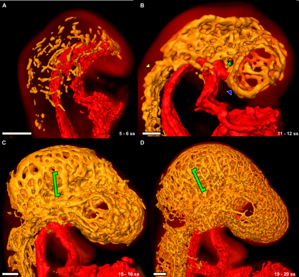
Figure 5. Development of the cephalic plexus between the 5 and 20 somite embryo.
(A) The vasculature in the 5 somite mouse embryo is a series of disconnected clusters of PECAM-1-expressing cells. The DA and the heart are surface rendered red, PECAM-1 expression throughout the cephalic mesenchyme is surface rendered orange, and the autofluorescence of the mouse embryo is volume rendered with a hot metal colourmap.
(B) By 11 somites, the cells have aggregated into a rudimentary vascular plexus. Larger vessels such as the PHV (blue arrowhead), the PMA (yellow arrowhead) and the ICA (green arrowhead) are visible (see also Supplemental Video S1). The PHV at this stage is a single large vessel that runs in an anterior-posterior direction starting from the cephalic flexure down to the first intersegmental vessel.
(C) The cephalic plexus has remodelled into a more stereotypic pattern by 15 somites. The cephalic veins are easily distinguishable (green bracket).
(D) At 19 somites the cephalic plexus has become more refined into recognizable structures. The cephalic veins are still visible at this stage (green bracket). All scale bars represent 100 microns.
Image Source: Three-Dimensional Analysis of Vascular Development in the Mouse Embryo Walls JR, Coultas L, Rossant J, Henkelman RM (2008) Three-Dimensional Analysis of Vascular Development in the Mouse Embryo. PLoS ONE 3(8): e2853. PLoS ONE
http://www.mouseimaging.ca/research/embryo_vasc_atlas.html
Citation: Walls JR, Coultas L, Rossant J, Henkelman RM (2008) Three-Dimensional Analysis of Vascular Development in the Mouse Embryo. PLoS ONE 3(8): e2853. doi:10.1371/journal.pone.0002853
Editor: Tailoi Chan-Ling, University of Sydney, Australia
Received: February 6, 2008; Accepted: June 11, 2008; Published: August 6, 2008
Copyright: © 2008 Walls et al. This is an open-access article distributed under the terms of the Creative Commons Attribution License, which permits unrestricted use, distribution, and reproduction in any medium, provided the original author and source are credited.
File history
Click on a date/time to view the file as it appeared at that time.
| Date/Time | Thumbnail | Dimensions | User | Comment | |
|---|---|---|---|---|---|
| current | 21:02, 11 October 2009 |  | 600 × 557 (559 KB) | S8600021 (talk | contribs) | Figure 5. Development of the cephalic plexus between the 5 and 20 somite embryo. (A) The vasculature in the 5 somite mouse embryo is a series of disconnected clusters of PECAM-1-expressing cells. The DA and the heart are surface rendered red, PECAM-1 exp |
You cannot overwrite this file.
File usage
There are no pages that use this file.
