File:Cardiac muscle EM02.jpg
Original file (1,072 × 735 pixels, file size: 224 KB, MIME type: image/jpeg)
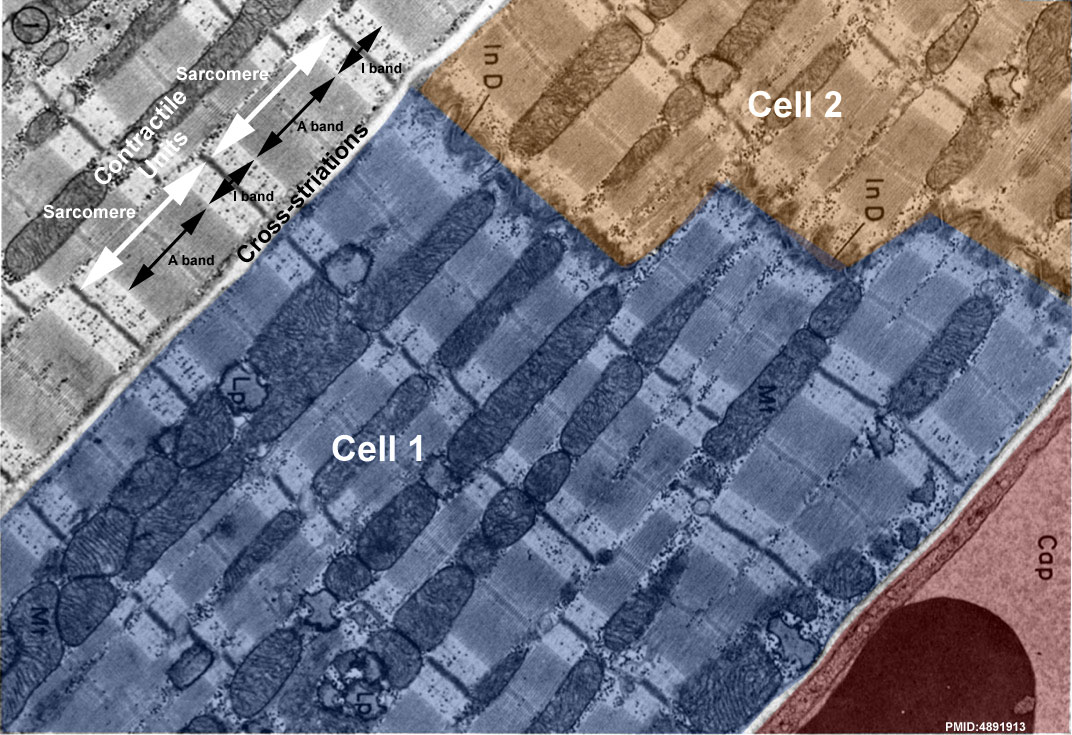
Cardiac Muscle Electron Micrograph
Electron micrograph of parts of three cat cardiac muscle fibers and an adjacent capillary in longitudinal section. This original historic (1969) EM has been relabeled to show key features in cardiac muscle ultrastructure. At the light microscope histology level, only the intercalated disc and some cross-striations can typically be seen using histological stains.
The cardiomyocytes (cardiac muscle cells) are packed with sarcomeres (for contraction) and mitochondria (for energy) with a nearby capillary for nutrient supply and waste removal.
- Image Top - In a cardiomyocyte (cardiac muscle cell) 2 contractile units (sarcomere) are shown by white arrows. The A and I bands, shown by black arrows, are the regions visible by light microscope as cross-striations.
- Image Bottom - The 2 cardiomyocytes (cardiac muscle cells) are coloured (labeled cell 1 and cell 2) and are joined by an intercalated disc.
- Image Bottom Right - A capillary (red) enclosed by an endothelial cell and its basement membrane contains a red blood cell.
Legend
- In D - Intercalated disc, the two lower cells are joined end to end by a typical steplike intercalated disc.
- Mt - Rows of mitochondria appear to divide the contractile substance into myofibril-like units but, unlike the true myofibrils of skeletal muscle, these branch and rejoin and are quite variable in width.
- Lp - Lipid droplets somewhat distorted in specimen preparation are found between the ends of the mitochondria.
- Cap - Capillary.
Original image X 15,000.
Reference
Fawcett DW & McNutt NS. (1969). The ultrastructure of the cat myocardium. I. Ventricular papillary muscle. J. Cell Biol. , 42, 1-45. PMID: 4891913
Copyright
Rockefeller University Press - Copyright Policy This article is distributed under the terms of an Attribution–Noncommercial–Share Alike–No Mirror Sites license for the first six months after the publication date (see http://www.jcb.org/misc/terms.shtml). After six months it is available under a Creative Commons License (Attribution–Noncommercial–Share Alike 4.0 Unported license, as described at https://creativecommons.org/licenses/by-nc-sa/4.0/ ). (More? Help:Copyright Tutorial)
Original article figure (FIG. 1) has been scaled and rotated. Labels and colours have also been added to the original image.
Cite this page: Hill, M.A. (2024, April 18) Embryology Cardiac muscle EM02.jpg. Retrieved from https://embryology.med.unsw.edu.au/embryology/index.php/File:Cardiac_muscle_EM02.jpg
- © Dr Mark Hill 2024, UNSW Embryology ISBN: 978 0 7334 2609 4 - UNSW CRICOS Provider Code No. 00098G
File history
Click on a date/time to view the file as it appeared at that time.
| Date/Time | Thumbnail | Dimensions | User | Comment | |
|---|---|---|---|---|---|
| current | 14:39, 6 August 2012 | | 1,072 × 735 (224 KB) | Z8600021 (talk | contribs) | |
| 14:35, 6 August 2012 | 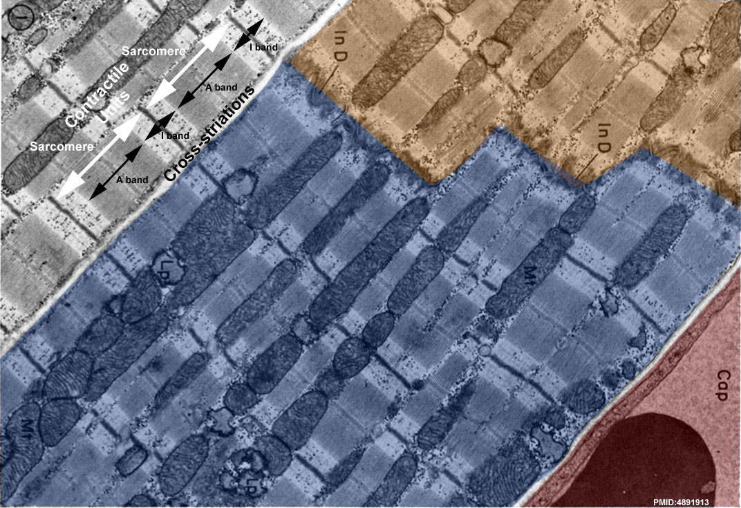 | 1,072 × 735 (222 KB) | Z8600021 (talk | contribs) | ==Cardiac Muscle Electron Micrograph== Electron micrograph of parts of three cat cardiac muscle fibers and an adjacent capillary in longitudinal section. This is a historic (1969) EM showing key features in cardiac muscle ultrastructure. Only the interca |
You cannot overwrite this file.