File:Canine oocyte to blastocyst.jpg
Original file (825 × 1,000 pixels, file size: 154 KB, MIME type: image/jpeg)
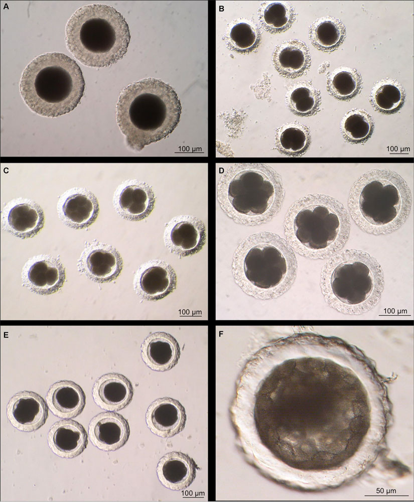
Canine Oocyte to Blastocyst
Canine oocytes and embryos observed under light microscopy at different stages after ovulation. Maturation stages were determined by confocal microscopy.
A Canine oocytes, Metaphase II, 82h (3.5 days) post-ovulation. The oocytes have lost their mucified mass, but are still surrounded by the corana radiata.
B Two-cell canine embryos collected 137h (5.7 days) after ovulation.
C Four-cell and eight-cell canine embryos, 153h (6.4 days) after ovulation.
D Eight-cell canine embryos collected 174h (7.2 days) after ovulation still have a few granulosa cells around their zona pellucida.
E Morula stage canine embryos collected 254h (10.6 days) after ovulation.
F Early blastocyst stage canine embryos, collected 265h (11 days) after ovulation.
Image Source: Image contributed by Dr Karine Reynaud, Department of Life Sciences and Health and UMR Developmental and Reproduction Biology ENVA, UMR 1198 Biologie du De ́veloppement et Reproduction, 7 Avenue du Ge ́ne ́ral De Gaulle, 94700 Maisons-Alfort, France. CINRA, UMR 1198 Biologie du De ́veloppement et Reproduction, F-78350 Jouy en Josas, France. Who kindly provided these images of the developing dog. Images are for educational purposes only and cannot be reproduced electronically or in writing without permission.
File history
Click on a date/time to view the file as it appeared at that time.
| Date/Time | Thumbnail | Dimensions | User | Comment | |
|---|---|---|---|---|---|
| current | 15:22, 12 April 2011 | | 825 × 1,000 (154 KB) | S8600021 (talk | contribs) | ==Canine Oocyte to Blastocyst== {{Template:Karine}} |
You cannot overwrite this file.
File usage
The following page uses this file: