File:Caldwell05.jpg

Original file (818 × 1,000 pixels, file size: 91 KB, MIME type: image/jpeg)
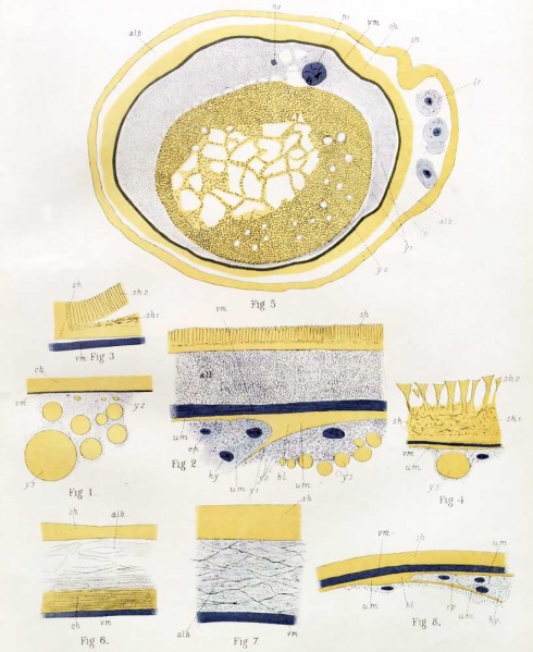
PLATE 30. Sections through Ornithorhynchus, Echidna and Phascolarctos
Fig. 1. Ornithorhynchus. - Small portion of a section through the segmenting ovum, taken from the open end of the Fallopian tube, measuring 2.6 mm. in diameter : ch, pro-albumen ; vm, vitelline membrane.
Fig. 2. Echidna. - Small portion of a section through the segmenting ovum taken from the uterus, and measuring G mm. in diameter : sh, shell ; alb, albumen ; vm, vitelline membrane ; ma, coagulum ; bl, blastopore ; ep, epiblast ; hy, hypoblast.
Fig. 3. Ornithorhynchus. - Small portion of a section through the segmenting ovum taken from the uterus, and measuring 6 mm. in diameter : sh, base of shell ; sh1, middle layer of ditto ; sh2, papillae of ditto.
Fig. 4. Echidna. - Small portion of a section through the blastodermic vesicle, taken from the uterus, and measuring 9 mm. in diameter : sh2 , cones derived from papilke of previous stage.
Fig. 5. Phascolarctos cinereus. - The 17th section of a vertical longitudinal series of 35 sections through the segmenting ovum, containing 2 nuclei, taken from the uterus, and measuring 0.17 mm. in diameter : sh, shell membrane; fe, cells of follicular epithelium ; alb, albumen ; ch, pro-albumen ; vm, vitelline membrane ; y t , protoplasm, with finest yolk granules ; y2, white yolk ; n, nucleus of smaller segmentation area ; n2, nucleus of larger segmentation area.
Fig. 6. Phascolarctos. - From uterus, 0.28 mm. in diameter, stage of 4 nuclei : sh, shell ; ch, pro-albumen ; alb, albumen ; vm, vitelline membrane.
Fig. 7. Phascolarctos. - From uterus, 0.31 mm. in diameter; lettering as in fig. G.
Fig. 8. Hypsiprymnus. - From uterus, 0.4 mm. in diameter: sh, shell membrane; vm, vitelline membrane ; um, coagulum ; bl, blastopore ; ep, epiblast ; hy, hypoblast. (Cf. fig. 2.)
Zeiss, oc. 2, obj. 1/18 homogen. : cam. luc.
| Historic Disclaimer - information about historic embryology pages |
|---|
| Pages where the terms "Historic" (textbooks, papers, people, recommendations) appear on this site, and sections within pages where this disclaimer appears, indicate that the content and scientific understanding are specific to the time of publication. This means that while some scientific descriptions are still accurate, the terminology and interpretation of the developmental mechanisms reflect the understanding at the time of original publication and those of the preceding periods, these terms, interpretations and recommendations may not reflect our current scientific understanding. (More? Embryology History | Historic Embryology Papers) |
- Links: plate 31 bw | plate 31
Reference
Caldwell WH. The Embryology of Monotremata and Marsupialia Part I. (1887) Phil. Trans. Roy. Soc 178 B.
Cite this page: Hill, M.A. (2024, April 19) Embryology Caldwell05.jpg. Retrieved from https://embryology.med.unsw.edu.au/embryology/index.php/File:Caldwell05.jpg
- © Dr Mark Hill 2024, UNSW Embryology ISBN: 978 0 7334 2609 4 - UNSW CRICOS Provider Code No. 00098G
File history
Click on a date/time to view the file as it appeared at that time.
| Date/Time | Thumbnail | Dimensions | User | Comment | |
|---|---|---|---|---|---|
| current | 10:52, 31 January 2012 |  | 818 × 1,000 (91 KB) | S8600021 (talk | contribs) | {{Caldwell1887}} {{Historic Disclaimer}} |
You cannot overwrite this file.
File usage
The following page uses this file:
