File:CNS later development.jpg
Original file (961 × 462 pixels, file size: 59 KB, MIME type: image/jpeg)
CNS Later Development
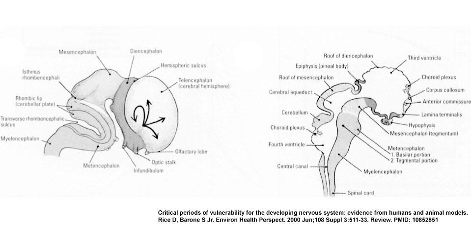
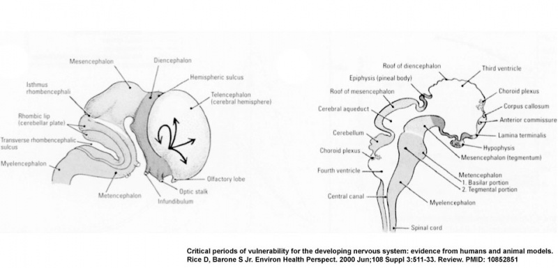
(E) The lateral view shows the migratory paths from the more central ventricular zone and gradients maturation of the neocortex (see arrows).
(F) The midsagittal view of the brain and spinal cord, with the major divisions delineated and the continuity of the ventricles noted; the formation of the choroid plexus corresponds to GD 13.5 in rats and GD 48-51 in humans.
Figure-3EF-PMID10852851.jpg
Image Source: Critical periods of vulnerability for the developing nervous system: evidence from humans and animal models. Rice D, Barone S Jr. Environ Health Perspect. 2000 Jun;108 Suppl 3:511-33. Review. PMID: 10852851 | Environmental Health Perspectives - Critical Periods of Vulnerability for the Developing Nervous System: Evidence from Humans and Animal Models | PMC: 1637807
File history
Click on a date/time to view the file as it appeared at that time.
| Date/Time | Thumbnail | Dimensions | User | Comment | |
|---|---|---|---|---|---|
| current | 01:07, 11 August 2009 | | 961 × 462 (59 KB) | MarkHill (talk | contribs) | CNS later development cartoon (E) The lateral view shows the migratory paths from the more central ventricular zone and gradients maturation of the neocortex (see arrows). (F) The midsagittal view of the brain and spinal cord, with the major divisions d |
You cannot overwrite this file.
File usage
There are no pages that use this file.