File:BurnsOgryzlo1938 fig05.jpg

Original file (790 × 1,000 pixels, file size: 97 KB, MIME type: image/jpeg)
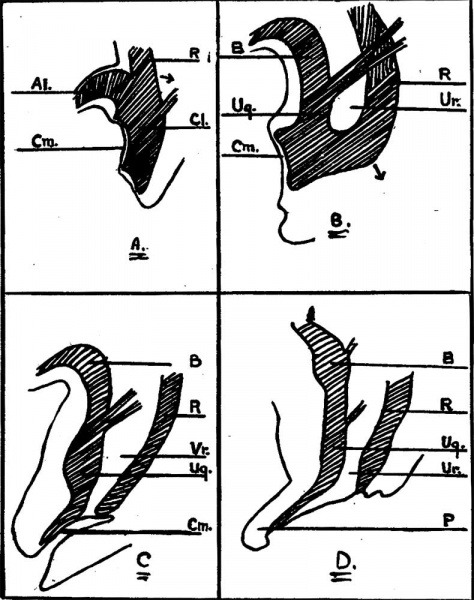
Fig. 5. Case 2. Differentiation of Cloaca
Al. - Allantois. B. — Bladder. cl.— cloaca. Cm.— Cloacal membrane. R. — Rectum. Ug. — Urogenital sinus. Ur. — Urorectal septum. P. — Penis.
Fig. 5a. In the early stages of development (4 weeks) the cloaca forms a triangle-shaped cavity at the tai1 end of the embryo, and receives the openings of the· allantois and rectum, as well as the Wolffian duct. 1ts ventral wall is largely formed by the cloacal membrane which extends upwards on the ventral wall of the allantois and on to the body stalk or umbilical cord. This membrane consists only of the two primitive layers, entoderm and ectoderm.
Fig. 5b. The cloacal orifice of the rectum then migrates backwards, leaving the ventral part of the cloaca to form the bladder and urogenital sinus, and gives the appearance of these two being separated from the rectum by a septum, called the urorectal septum.
Fig. 5c. When the recta1 orifice reaches the anal depressions it becomes separated from the urogenital sinus.
Fig. 5d. The urorectal septum in the male gives rise to the greater part of the floor of the penile urethra and perineum, while in the female the uterus and vagina develop in it.
| Historic Disclaimer - information about historic embryology pages |
|---|
 |
Reference
Burns CW. and Ogryzlo MA. Congenital hernia into the umbilical cord; two cases, one associated with persistent cloaca. (1938) Can Med Assoc J. 39(5): 438-41. PMID 20321146
Cite this page: Hill, M.A. (2024, April 16) Embryology BurnsOgryzlo1938 fig05.jpg. Retrieved from https://embryology.med.unsw.edu.au/embryology/index.php/File:BurnsOgryzlo1938_fig05.jpg
- © Dr Mark Hill 2024, UNSW Embryology ISBN: 978 0 7334 2609 4 - UNSW CRICOS Provider Code No. 00098G
File history
Click on a date/time to view the file as it appeared at that time.
| Date/Time | Thumbnail | Dimensions | User | Comment | |
|---|---|---|---|---|---|
| current | 10:31, 22 August 2018 | | 790 × 1,000 (97 KB) | Z8600021 (talk | contribs) | |
| 10:28, 22 August 2018 |  | 925 × 1,334 (140 KB) | Z8600021 (talk | contribs) |
You cannot overwrite this file.
File usage
The following page uses this file: