File:BurgosFawcett1955 fig11.jpg

Original file (1,453 × 2,015 pixels, file size: 528 KB, MIME type: image/jpeg)
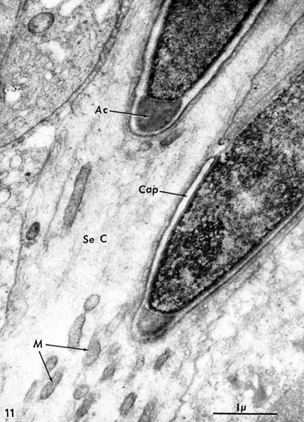
Fig. 11. A Vertical section through a Sertoli cell
Including part of two late spermatids and showing the intimate relation of the developing heads to the surrounding Sertoli cell. Observe the beginning condensation of the nuclear material into coarse osmiophilic granules, and the clustering of Sertoli cell mitochondria around the head of the spermatid. X 25,699.
Reference
Burgos MH. and Fawcett DW. Studies on the Fine Structure of the Mammalian Testis I. Differentiation of the Spermatids in the Cat (Felis Domesrica) (1955) J. Biophxsic. Biochem. Cytol. 1(4): 287-300. PMID 13242594
Copyright
Rockefeller University Press - Copyright Policy This article is distributed under the terms of an Attribution–Noncommercial–Share Alike–No Mirror Sites license for the first six months after the publication date (see http://www.jcb.org/misc/terms.shtml). After six months it is available under a Creative Commons License (Attribution–Noncommercial–Share Alike 4.0 Unported license, as described at https://creativecommons.org/licenses/by-nc-sa/4.0/ ). (More? Help:Copyright Tutorial)
Cite this page: Hill, M.A. (2024, April 20) Embryology BurgosFawcett1955 fig11.jpg. Retrieved from https://embryology.med.unsw.edu.au/embryology/index.php/File:BurgosFawcett1955_fig11.jpg
- © Dr Mark Hill 2024, UNSW Embryology ISBN: 978 0 7334 2609 4 - UNSW CRICOS Provider Code No. 00098G
File history
Click on a date/time to view the file as it appeared at that time.
| Date/Time | Thumbnail | Dimensions | User | Comment | |
|---|---|---|---|---|---|
| current | 21:16, 30 May 2018 |  | 1,453 × 2,015 (528 KB) | Z8600021 (talk | contribs) | |
| 21:13, 30 May 2018 |  | 1,611 × 2,393 (545 KB) | Z8600021 (talk | contribs) |
You cannot overwrite this file.
File usage
The following page uses this file: