File:Buell10.jpg

Original file (665 × 1,000 pixels, file size: 113 KB, MIME type: image/jpeg)
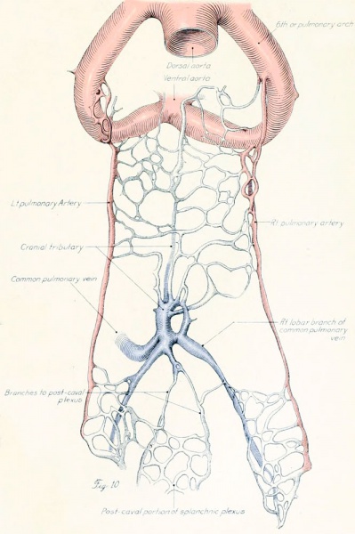
Fig. 10. Origin of the Pulmonary Vessels in the Chick
Dissections of injected chick embryos of 85 hours' incubation. Figure 9 shows the right side of the pulmonary system. In figure 10 the spinal cord, dorsal aorta, and dorsal surface of the gut have been removed, exposing the pulmonary system in a coronal plane from a dorsal view. The lung is in a simple stage of right and left primary buds which do not show further lobulation. The left bud is more ventral than the right and is parallel to the gut. The right bud tends more toward a horizontal position in relation to the plane of the gut. The pulmonary vessels bear a constant relation to the bronchi of the buds, even at this early stage. The artery lies dorsal and lateral to the bronchus; the vein, ventral and medial to the bronchus, the lung capillaries lying between the two on the dorsal surface of the buds. The pulmonary artery comes off from the arch at the junction of its middle and proximal third, and passes directly back to the tip of the lung-bud, where it joins freely, in a capillary net, with the corresponding tributary of the pulmonary vein. Very near the arch a capillary connection is given off to the anterior cardinal vein. The two arteries extend parallel to each other and in their proximal third are joined by numerous capillary anastomoses which are drained by the cranial tributary of the common vein. The middle third of the artery has no branches. The entire distal third is connected with the vein by a rich plexus of capillaries over the dorsal surface of the lung-bud. A few twigs are still present, connecting with the post-caval portion of the plexus. The pulmonary vein is made up of several tributaries which unite in a common trunk; this in turn empties into the sinus venosus. Considerable variation is encountered in the pattern of these branches in different specimens. The right and left lobar branches to the lung-buds drain their respective arteries. In figure 9 a vessel connects the right lobar vein to the cranial tributary. This is not constant and is absent in figure 10. A few small branches to the post-caval plexus are seen caudal to the lobar branches. The cranial tributary of the common vein drains the anastomotic vessels between the two pulmonary arteries and arches. It extends directly caudad on the ventral surface of the gut and, with the other tributaries, empties into the common vein. It may have but one opening into the common vein, as in figure 10. This stage is about the oldest in which the cranial tributary is seen complete and represents its highest development. In a later stage, as described by Squier, the cranial tributary loses its arterial connections and disappears. The pulmonary arches (sixth) have undergone rapid growth and have included the arteries within their walls.
- 1922 Chicken Pulmonary: Fig 1 | Fig 2 | Fig 3 | Fig 4 | Fig 5 | Fig 6 | Fig 7 | Fig 8 | Fig 9 | Fig 10 | Plate 1 | Plate 2 | Carnegie No.66 | Chicken Development | Respiratory
- Links: Carnegie Institution of Washington - Contributions to Embryology | Chicken Development | Respiratory System Development
| Historic Disclaimer - information about historic embryology pages |
|---|
 |
File history
Click on a date/time to view the file as it appeared at that time.
| Date/Time | Thumbnail | Dimensions | User | Comment | |
|---|---|---|---|---|---|
| current | 16:23, 23 January 2013 | | 665 × 1,000 (113 KB) | Z8600021 (talk | contribs) | JP2 figure scan |
| 12:25, 29 March 2011 |  | 621 × 966 (83 KB) | S8600021 (talk | contribs) | ==Origin of the Pulmonary Vessels in the Chick - Fig. 10=== {{Template:Historic Disclaimer}} ===Figs. 9-10=== Dissections of injected chick embryos of 85 hours' incubation. Figure 9 shows the right side of the pulmonary system. In figure 10 the spinal |
You cannot overwrite this file.
File usage
The following page uses this file: