File:Buell03.jpg
From Embryology

Size of this preview: 592 × 599 pixels. Other resolution: 790 × 800 pixels.
Original file (790 × 800 pixels, file size: 73 KB, MIME type: image/jpeg)
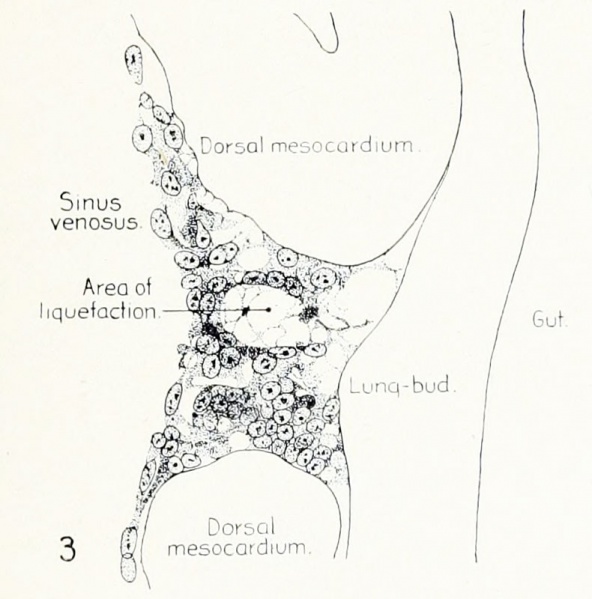
Fig. 3. Sagittal section (10 micron) through left third of chick of 23 somites
Sagittal section (10 micron) through left third of proliferation of angioblasts, to show process of liquefaction extending toward sinus venosus. It is about to open into the sinus, thus forming the common pulmonary vein. Hematoxylin and eosin stain; chick of 23 somites, 48 hours' incubation, series D.
- 1922 Chicken Pulmonary: Fig 1 | Fig 2 | Fig 3 | Fig 4 | Fig 5 | Fig 6 | Fig 7 | Fig 8 | Fig 9 | Fig 10 | Plate 1 | Plate 2 | Carnegie No.66 | Chicken Development | Respiratory
- Links: Carnegie Institution of Washington - Contributions to Embryology | Chicken Development | Respiratory System Development
| Historic Disclaimer - information about historic embryology pages |
|---|
 |
File history
Click on a date/time to view the file as it appeared at that time.
| Date/Time | Thumbnail | Dimensions | User | Comment | |
|---|---|---|---|---|---|
| current | 18:13, 23 January 2013 | | 790 × 800 (73 KB) | Z8600021 (talk | contribs) | {{Buell1922}} :'''Links:''' Carnegie Institution of Washington - Contributions to Embryology | Chicken Development | Respiratory System Development {{Historic Disclaimer}} Category:Cardiovascular [[ |
You cannot overwrite this file.
File usage
The following page uses this file: