File:Bryce1908 plate10.jpg
From Embryology
Size of this preview: 450 × 599 pixels. Other resolution: 1,100 × 1,465 pixels.
Original file (1,100 × 1,465 pixels, file size: 184 KB, MIME type: image/jpeg)
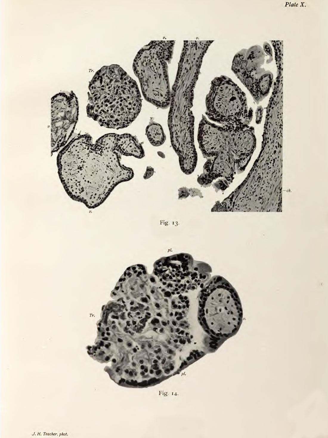
Plate 10. Chorionic Villi sections
Fig. 13. Section of the Villi and Edge of the Chorion. Photograph. x 140 d.
The section demonstrates that the villi are covered l>y a double- layered epithelium (cellular layer and plasiuodial layer).
Fig. 14. Section of a Single Villus and Mass of Lanohans' Layer Cells, more higlily magnified than in the last figure. Photogmph. x 240 d.
The two-layered epithelium covering the mesoblastic core of the villus is well seen. The mass of Langhans' layer cells is covered by a very distinct plasmodial layer (syncytium of authors).
- Bryce 1908 Human Ovum: Plate 1 | Plate 2 | Plate 3 | Plate 4 | Plate 5 | Plate 6 | Plate 7 | Plate 8 | Plate 9 | Plate 10
| Historic Disclaimer - information about historic embryology pages |
|---|
 |
File history
Click on a date/time to view the file as it appeared at that time.
| Date/Time | Thumbnail | Dimensions | User | Comment | |
|---|---|---|---|---|---|
| current | 08:57, 3 November 2013 | | 1,100 × 1,465 (184 KB) | Z8600021 (talk | contribs) | ==Plate 10.== |
You cannot overwrite this file.
File usage
The following page uses this file: