File:Bryce1908 plate09.jpg
Original file (1,203 × 1,000 pixels, file size: 115 KB, MIME type: image/jpeg)
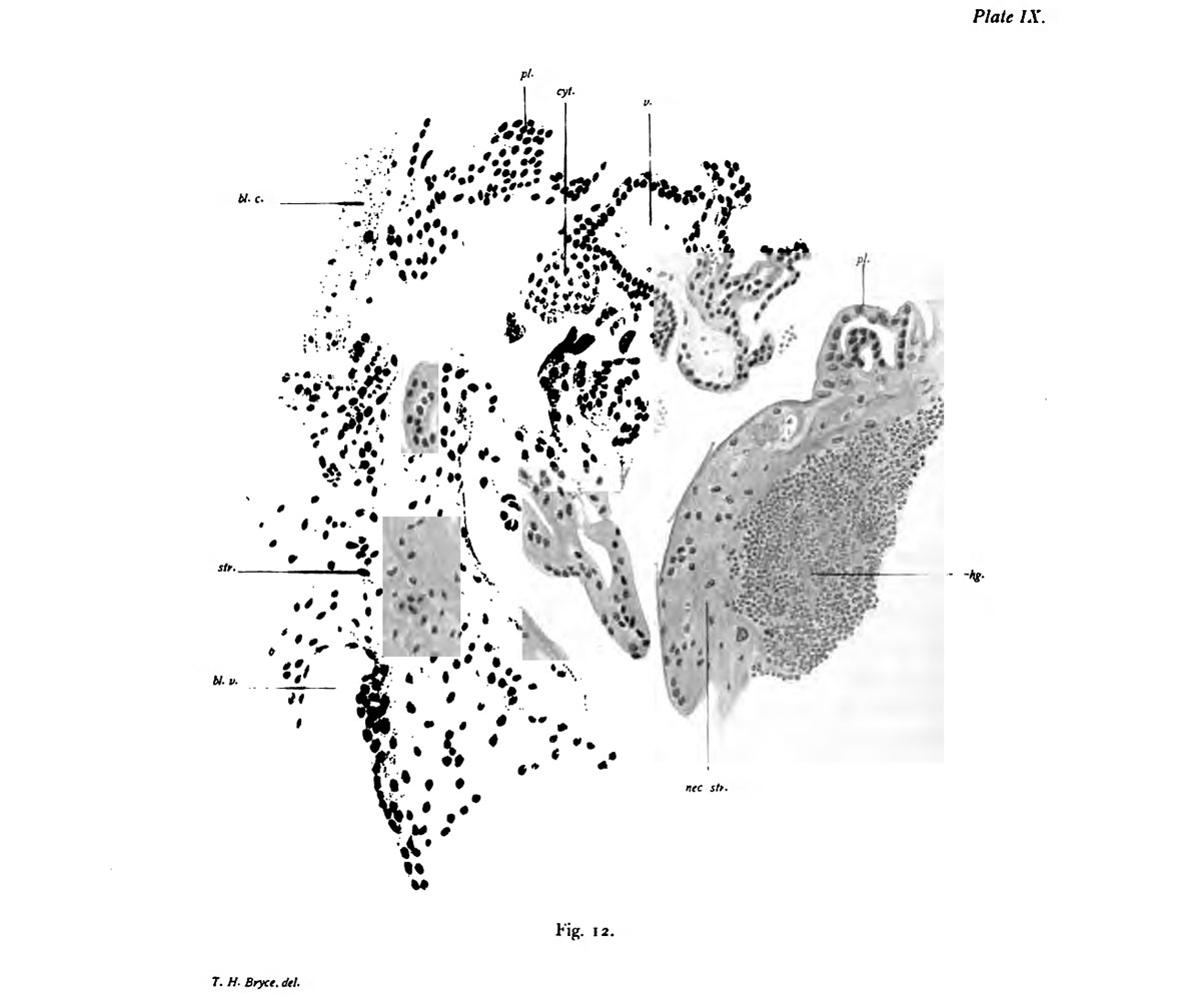
Plate 9. Section of a portion of the ovarian stroma and villi of the chorion
Fig. 12. Section of a portion of the ovarian stroma and villi of the chorion x200 d.
r, villus ; 7r.j mass of Langhans' layer cells ; pl, plasmodium ; bl.c., lilood clot ; hg., haemorrage ; sfr., stroma of ovary; bl.e., hlood-vessel in stroma.
A portion of the stroma lias been selected in which runs a large vessel ; this, wlien traced thniugh the sections, was found to o|)en out into the gest«ition sjic The stroma adjoining the wivity is necrotic. Masses of pliusmodium are .seen attached to it, ImiIIi on the right ahove the haemorrhage, and to the left alK)ve the blood- vessel.
- Bryce 1908 Human Ovum: Plate 1 | Plate 2 | Plate 3 | Plate 4 | Plate 5 | Plate 6 | Plate 7 | Plate 8 | Plate 9 | Plate 10
| Historic Disclaimer - information about historic embryology pages |
|---|
 |
File history
Click on a date/time to view the file as it appeared at that time.
| Date/Time | Thumbnail | Dimensions | User | Comment | |
|---|---|---|---|---|---|
| current | 08:54, 3 November 2013 | | 1,203 × 1,000 (115 KB) | Z8600021 (talk | contribs) |
You cannot overwrite this file.
File usage
The following page uses this file: