File:Brewer1937 plate07.jpg

Original file (1,280 × 1,713 pixels, file size: 444 KB, MIME type: image/jpeg)
Plate 7
14 This photomicrograph of the decidua. Vera shows the enlarged and tortuous glands filled with secretion. The superficial venous sinuses are dilated. The spiral arteries extend to the surface of the vera. The superficial half of the Vera is edematous. Surrounding the spiral arteries the deeidual tissues are more dense. x 54.1.
15 The distal portion of the spiral artery entering the penetration zone is filled with blood. The proximal portion (not shown in the photograph) contains no blood and is constricted. The artery ends as an endothelial tube in the penetration zone. See figures 18 and 19. X 32.4.
16 A spiral artery in the lateral penetration zone divides into two branches. One branch is filled with blood and the other is constricted and empty. X 68.
17 The gland epithelium with syncytial elements adjacent is destroyed in places. A capillary has been disrupted and its free end projects into the gland lumen. The latter is filled with blood. X 395.
| Historic Disclaimer - information about historic embryology pages |
|---|
 |
- Links: plate 1 | fig 1 | fig 2 | fig 3 | plate 2 | fig 4 | plate 3 | fig 5 | fig 6 | plate 4 | fig 7 | fig 8 | fig 9 | fig 10 | plate 5 | plate 6 | plate 7 | plate 8 | plate 9 | plate 10 | plate 11 | plate 12 | plate 13 | plate 14 | fig 11 | 1937 Brewer | Historic Papers | Carnegie Embryo 8819 | Carnegie stage 6 | Week 2
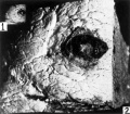
Fig 1 and 2 Tissue block removed
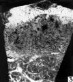
Fig 3 Hemorrhagic region
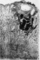
Fig 4 Blastocyst directly beneath surface hemorrhage
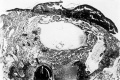
Fig 5 Blastocyst entirely surrounded by short villi
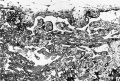
Fig 6 Portion of the chorionic wall shows two short villi
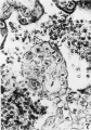
Fig 7 Cytotrophoblastic cell mitosis
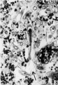
Fig 8 Fetal cell is shown amid maternal tissue
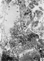
Fig 9 Lateral sinus and fetal trophoblast
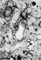
Fig 10 Spiral arteriole with a syncytial cell
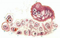
Fig 11 Drawing of cytotrophoblastic cells
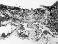
Fig 12 Operculum with one lateral margin
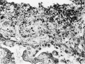
Fig 13 Operculum with adherent villus
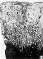
Fig 14 Decidua
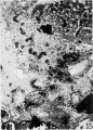
Fig 15 Distal portion of the spiral artery
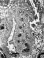
Fig 16 Spiral artery in the lateral penetration zone
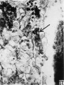
Fig 17 Gland epithelium with syncytial elements
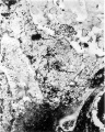
Fig 18 A spiral artery with hemorrhage
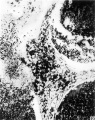
Fig 19 End of this spiral artery in penetration zone
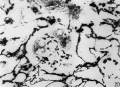
Fig 20 Reticulum in the maternal tissue
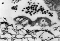
Fig 21 Junction of the maternal and fetal tissues
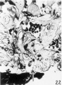
Fig 22 Wall of an endometrial gland
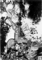
Fig 23 Endometrial gland
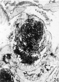
Fig 24 Endometrial gland in implantation cavity
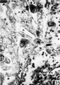
Fig 25 Surface epithelium of the decidua
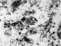
Fig 26 Trophoblastic cell
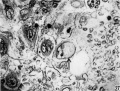
Fig 27 Trophoblastic cell
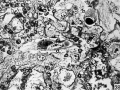
Fig 28 Trophoblastic cell
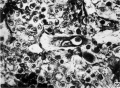
Fig 29 Trophoblastic cell phagocytized lymphoyte
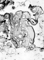
Fig 30 Multinucleated syncytial mass


Reference
Brewer JI. A normal human ovum in a stage preceding the primitive streak (The Edwards-Jones-Brewer ovum). (1937) Amer. J Anat., 61: 429-481.
Cite this page: Hill, M.A. (2024, April 24) Embryology Brewer1937 plate07.jpg. Retrieved from https://embryology.med.unsw.edu.au/embryology/index.php/File:Brewer1937_plate07.jpg
- © Dr Mark Hill 2024, UNSW Embryology ISBN: 978 0 7334 2609 4 - UNSW CRICOS Provider Code No. 00098G
File history
Click on a date/time to view the file as it appeared at that time.
| Date/Time | Thumbnail | Dimensions | User | Comment | |
|---|---|---|---|---|---|
| current | 12:37, 3 February 2017 | | 1,280 × 1,713 (444 KB) | Z8600021 (talk | contribs) | |
| 11:59, 3 February 2017 |  | 1,593 × 2,259 (626 KB) | Z8600021 (talk | contribs) | ===Plate 7=== 14 This photomicrograph of the decidua. Vera shows the enlarged and tortuous glands filled with secretion. The superficial venous sinuses are dilated. The spiral arteries extend to the surface of the vera. The superficial half of the Ver... |
You cannot overwrite this file.
File usage
The following page uses this file: