File:Brewer1937 plate03.jpg

Original file (1,583 × 2,260 pixels, file size: 597 KB, MIME type: image/jpeg)
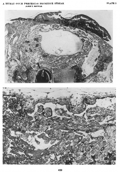
Plate 3
5 The blastocyst is entirely surrounded by short villi. The villi are most numerous on the basal side. The embryonic anlage is attached to the most dependent pole. The amnion is larger than the yolk sac. The embryonic plate is a simple, flat structure without distinctive surface markings. There is a condensation of cells in the region of the body stalk. The large basal and lateral. sinuses surround the blastocyst almost completely. The coagulum on the surface overlies the surface epithelium. It consists of blood and some fetal and maternal cells. The depression in the central portion is the point of entrance. Section 23-1-4. x 27.5.
6 This portion of the chorionic wall shows two short villi. with mesodermal cores. The Langhans cells extend out beyond the entl of the villus and join with those from the neighboring villi. The syncytiiun resembles the so-called primary syncytiunl. Maternal blood is found in the spaces between the fetal tissues. X 180.8.
| Historic Disclaimer - information about historic embryology pages |
|---|
 |
- Links: plate 1 | fig 1 | fig 2 | fig 3 | plate 2 | fig 4 | plate 3 | fig 5 | fig 6 | plate 4 | fig 7 | fig 8 | fig 9 | fig 10 | plate 5 | plate 6 | plate 7 | plate 8 | plate 9 | plate 10 | plate 11 | plate 12 | plate 13 | plate 14 | fig 11 | 1937 Brewer | Historic Papers | Carnegie Embryo 8819 | Carnegie stage 6 | Week 2
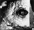
Fig 1 and 2 Tissue block removed
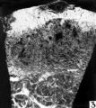
Fig 3 Hemorrhagic region
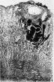
Fig 4 Blastocyst directly beneath surface hemorrhage
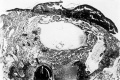
Fig 5 Blastocyst entirely surrounded by short villi
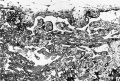
Fig 6 Portion of the chorionic wall shows two short villi
Fig 7 Cytotrophoblastic cell mitosis
Fig 8 Fetal cell is shown amid maternal tissue
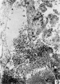
Fig 9 Lateral sinus and fetal trophoblast
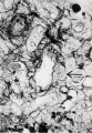
Fig 10 Spiral arteriole with a syncytial cell
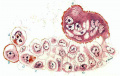
Fig 11 Drawing of cytotrophoblastic cells
Fig 12 Operculum with one lateral margin
Fig 13 Operculum with adherent villus
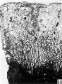
Fig 14 Decidua
Fig 15 Distal portion of the spiral artery
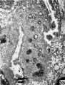
Fig 16 Spiral artery in the lateral penetration zone
Fig 17 Gland epithelium with syncytial elements
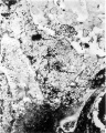
Fig 18 A spiral artery with hemorrhage
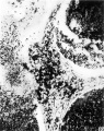
Fig 19 End of this spiral artery in penetration zone
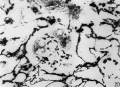
Fig 20 Reticulum in the maternal tissue
Fig 21 Junction of the maternal and fetal tissues
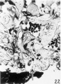
Fig 22 Wall of an endometrial gland
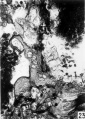
Fig 23 Endometrial gland
Fig 24 Endometrial gland in implantation cavity
Fig 25 Surface epithelium of the decidua
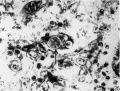
Fig 26 Trophoblastic cell
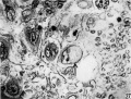
Fig 27 Trophoblastic cell
Fig 28 Trophoblastic cell
Fig 29 Trophoblastic cell phagocytized lymphoyte
Fig 30 Multinucleated syncytial mass


Reference
Brewer JI. A normal human ovum in a stage preceding the primitive streak (The Edwards-Jones-Brewer ovum). (1937) Amer. J Anat., 61: 429-481.
Cite this page: Hill, M.A. (2024, April 25) Embryology Brewer1937 plate03.jpg. Retrieved from https://embryology.med.unsw.edu.au/embryology/index.php/File:Brewer1937_plate03.jpg
- © Dr Mark Hill 2024, UNSW Embryology ISBN: 978 0 7334 2609 4 - UNSW CRICOS Provider Code No. 00098G
File history
Click on a date/time to view the file as it appeared at that time.
| Date/Time | Thumbnail | Dimensions | User | Comment | |
|---|---|---|---|---|---|
| current | 10:57, 3 February 2017 | | 1,583 × 2,260 (597 KB) | Z8600021 (talk | contribs) |
You cannot overwrite this file.
File usage
The following page uses this file: