File:Boyden1931 fig02.jpg

Original file (773 × 860 pixels, file size: 75 KB, MIME type: image/jpeg)
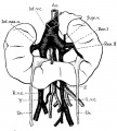
Fig. 2. Horseshoe kidney
Horseshoe kidney of seven-year-old girl, associated with double vena cava that lies posterior to kidneys; representing persistence of right and left supmoardinal veins of embryo. (After Marzynski, case 3; legends by E. A. B.). Note fetal lobulations of kidney.
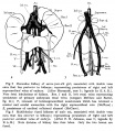
Ren. I and II, left renal veins representing persistence of primary embryonic renal veins (compare McClure and Butler, fig. 14); Y, remnant of intersupracardinal anastomosis which has retained a. cranial and caudal connection with the right supracardinal vein (McClure); Z, persistence of cardinal collateral channel (McClure).
Reference
Boyden EA. Description of a horseshoe kidney associated with left inferior vena cava and disc-shaped suprarenal glands, together with a note on the occurrence of horseshoe kidneys in human embryos. (1931) Anat. Rec. 51(2): 187-211.
Cite this page: Hill, M.A. (2024, April 25) Embryology Boyden1931 fig02.jpg. Retrieved from https://embryology.med.unsw.edu.au/embryology/index.php/File:Boyden1931_fig02.jpg
- © Dr Mark Hill 2024, UNSW Embryology ISBN: 978 0 7334 2609 4 - UNSW CRICOS Provider Code No. 00098GCatgeory:1930's
File history
Click on a date/time to view the file as it appeared at that time.
| Date/Time | Thumbnail | Dimensions | User | Comment | |
|---|---|---|---|---|---|
| current | 10:45, 8 September 2017 |  | 773 × 860 (75 KB) | Z8600021 (talk | contribs) | |
| 10:45, 8 September 2017 |  | 1,360 × 1,561 (325 KB) | Z8600021 (talk | contribs) |
You cannot overwrite this file.
File usage
The following 2 pages use this file: