File:Bonnot1906 fig01.jpg
From Embryology
Size of this preview: 626 × 599 pixels. Other resolution: 658 × 630 pixels.
Original file (658 × 630 pixels, file size: 61 KB, MIME type: image/jpeg)
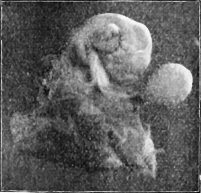
Fig. 1. From a photograph of the 11 mm human embryo (No. H60)
Somewhat magnified, showing the right side of the embryo, with the umbilical cord, yolk sac, and membranes. The tip of the tail is turned somewliat to the left, and is hidden by the umbilical cord.
| Week: | 1 | 2 | 3 | 4 | 5 | 6 | 7 | 8 |
| Carnegie stage: | 1 2 3 4 | 5 6 | 7 8 9 | 10 11 12 13 | 14 15 | 16 17 | 18 19 | 20 21 22 23 |
Reference
Bonnet E. and Severs R. On the structure of a human embryo eleven millimeters in length. (1906) Anat. Anz., 29: 452-459.
Cite this page: Hill, M.A. (2024, April 25) Embryology Bonnot1906 fig01.jpg. Retrieved from https://embryology.med.unsw.edu.au/embryology/index.php/File:Bonnot1906_fig01.jpg
- © Dr Mark Hill 2024, UNSW Embryology ISBN: 978 0 7334 2609 4 - UNSW CRICOS Provider Code No. 00098G
File history
Click on a date/time to view the file as it appeared at that time.
| Date/Time | Thumbnail | Dimensions | User | Comment | |
|---|---|---|---|---|---|
| current | 16:49, 19 December 2016 | | 658 × 630 (61 KB) | Z8600021 (talk | contribs) |
You cannot overwrite this file.
File usage
The following page uses this file: