File:Blyakher1955 fig07.jpg

Original file (1,028 × 1,482 pixels, file size: 178 KB, MIME type: image/jpeg)
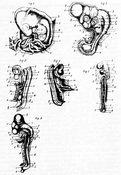
Fig. 7. Table II illustration from Wolff's "On the Formation of the Intestine"
1. Embryo of 4 days incubation; r - excised part of the vascular area on which the vesicle lies; a, b, c - vesicle or false amnion; g, h, i, 1- - translucent embryo; h - occiput;
g - anterior part of the head; i - vertebra; 1 - left wing; k - left auricle; B - left lateral part of thorax; C - fold of abdominal opening and true amnion (Table I , Fig. 2, ii, Fig. 7, xx) ; e - intestinal fold and fold of false amnion (see Table I, Fig. 7, n) ; m - opening between true and false amnion; d - part of middle intestine; f - rounded fossa, appearing from cardiac fossa, groove and lower fossa; N - lower fossa; p - right yolk vein; o - artery of that side; n - left yolk artery; q- -umbilical vesicle (allantois) .
2. The same object with extracted false amnion; a - part of upper layer of vascular area; b- -true amnion; c - occiput; d - anterior part of the head; D- middle protuberance, disappearing after six days; e- -vertebra; f - left wing;
F - left leg; g - left auricle; h- - lateral part of thorax; i - folds of abdominal opening from which membrane of true amnion takes origin; H, H- -the area of the abdomen, adjacent to the thighs, forming paired cavities in the sides of the vertebrae; E- - backwards-bending part of this cavity; k, 1, m, n, o, p- parts beginning from abdominal cavity; k - intestinal fold from which the membrane of the false amnion continues (n) ; 1 - deeper fold of intestine; m - parts of folded middle intestine; n - part of false amnion; o - its part continuing through the cardiac fossa to the stomach; p- upper part of the rectum.
3. The same object; amnion destroyed, lateral parts of thorax removed; and also abdominal cavity, extremities and head; viscera is visible; a - left chamber of heart; b - aural canal; c - left auricle; d - arch of aorta; v- - venous sinus; f - left lobe of liver; g - rudiment of abdomen; h - digestive tract; i - stomach; k - intestinal fold; 1 - duodenum; MM - middle intestine; n - part of membrane of false amnion; o - part of this membrane covering the intestinal canal; p- upper funnel-shaped part of the rectum; q - lower part of rectum, which, like the stomach, also already has cylindrical form, while lying between them a part of intestine opened; r - mesentery - continuation of false amnion and intestinal membranes; s - left kidney; t - part of vertebrae; u - left yolk artery.
4. The same preparation from the right side; a- - left chamber of the heart; b - its right chamber; c - aorta; d - arch of aorta; e - rudiment of left lung; f - right lobe of liver;
g - right kidney; h - right yolk artery; i - right intestinal membrane, from which middle intestine is formed; k - rectum; 1- mesentery; m - right membrane of false amnion; n - right yolk vein; N - trunk of vein; o - vertebrae.
5. Embryo represented in Table 1, Fig. 6, liberated from all membrane; a - anterior part of the head; b- - occiput; c, d - occipital region; d, e - region of thorax; e-z- the rest of the vertebrae; g - left part of the lower jaw; f - -process (first rib) ; h - left auricle; i - aural canal; k - left chamber of heart; 1 - aorta; m - membrane represented in Fig. 3, g; n - part of false amnion from which the cardiac fossa and stomach are formed; o, p, q, s, t, z - first rudiment of abdomen in the form of bending membrane; o - upper part of this membrane; p, s- edge of abdominal fold; q - its upper left, strongly curved part; t - its edge in the place where it becomes wider; v - right lateral abdominal membrane; u - left membrane; s, r, z - intestinal canal (fistula intestinalis) - first rudiment of mesentery with still divided membranes; s - right mesenterical membrane with right kidney; r - the same also - left; w- - opening between membranes of mesentery, in which uncovered vertebra is seen; x - vertebrae; y - rudiment of tailbone; z - rudiment of the pelvis .
6 . Embryo represented in Table I , Fig . 7 ; a - anterior part of the head; b - posterior part of the head; c - protuberance (see Fig. 2, D) ; d - thoracic part of the vertebrae; e - occipital region; f - rudiments of ribs; g - rudiment of wing; h - rudiment of leg; i - hip region; k - region of tailbone; 1 - rudiment of pelvic cavity; m - rudiment of true amnion; n - part of thorax; o - left auricle; q- - middle chamber; r a bdomen ; s - stomach; t - ascending vein; u - part of cephalic branch; v - edge of the stomach (see Table I, Fig. 1 , k) -, w - intestinal fold; x - part of false amnion; y - completely formed mesentery; z - upper, funnel-shaped part of the rectum; A - left, kidney, later on separated from the mesentery; B - yolk artery.
| Historic Disclaimer - information about historic embryology pages |
|---|
| Pages where the terms "Historic" (textbooks, papers, people, recommendations) appear on this site, and sections within pages where this disclaimer appears, indicate that the content and scientific understanding are specific to the time of publication. This means that while some scientific descriptions are still accurate, the terminology and interpretation of the developmental mechanisms reflect the understanding at the time of original publication and those of the preceding periods, these terms, interpretations and recommendations may not reflect our current scientific understanding. (More? Embryology History | Historic Embryology Papers) |
Figures: 1 Arnold van der Hulst | 2 Kaspar Friedrich Wolff | Wolff's chicken development
Reference
Blyakher L. History of embryology in Russia from the middle of the eighteenth to the middle of the nineteenth century (istoryia embriologii v Rossii s serediny XVIII do serediny XIX veka) (1955) Academy of Sciences USSR. Institute of the History of Science and Technology. Translation Smithsonian Institution (1982).
Cite this page: Hill, M.A. (2024, April 20) Embryology Blyakher1955 fig07.jpg. Retrieved from https://embryology.med.unsw.edu.au/embryology/index.php/File:Blyakher1955_fig07.jpg
- © Dr Mark Hill 2024, UNSW Embryology ISBN: 978 0 7334 2609 4 - UNSW CRICOS Provider Code No. 00098G
File history
Click on a date/time to view the file as it appeared at that time.
| Date/Time | Thumbnail | Dimensions | User | Comment | |
|---|---|---|---|---|---|
| current | 22:46, 10 May 2018 |  | 1,028 × 1,482 (178 KB) | Z8600021 (talk | contribs) | |
| 22:44, 10 May 2018 |  | 2,193 × 3,063 (347 KB) | Z8600021 (talk | contribs) | Figure 7. Table II illustration from Wolff's "On the Formation of the Intestine . " {{Blyakher1955 figures}} |
You cannot overwrite this file.
File usage
The following 2 pages use this file:
