File:Beattie1924 fig02.jpg

Original file (1,000 × 539 pixels, file size: 194 KB, MIME type: image/jpeg)
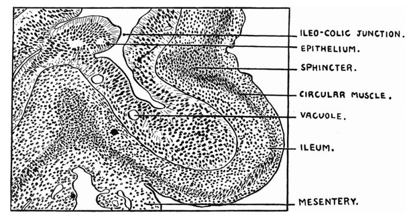
Fig. 2. The ileo-colic junction in a 32 mm human embryo
( x 200)
In the 32 mm. stage the ileo-colic angle has become a right angle, the terminal ileum now entering the colon from above and on the right. There is now at the junction (fig. 2) a well marked ring of muscle tissue which has invaded the sub-mucous layer of the gut and reduced it at the junction to a thin layer of cells; the outer or sub-serous layer remains of the same thickness as in the terminal ileum and proximal colon.
Reference
Beattie J. The early stages of the development of the ileo-colic sphincter. (1924) J Anat. 59: 56-59. PMID 17104039
Cite this page: Hill, M.A. (2024, April 24) Embryology Beattie1924 fig02.jpg. Retrieved from https://embryology.med.unsw.edu.au/embryology/index.php/File:Beattie1924_fig02.jpg
- © Dr Mark Hill 2024, UNSW Embryology ISBN: 978 0 7334 2609 4 - UNSW CRICOS Provider Code No. 00098G
File history
Click on a date/time to view the file as it appeared at that time.
| Date/Time | Thumbnail | Dimensions | User | Comment | |
|---|---|---|---|---|---|
| current | 19:45, 2 March 2020 |  | 1,000 × 539 (194 KB) | Z8600021 (talk | contribs) | |
| 19:37, 2 March 2020 |  | 1,422 × 782 (240 KB) | Z8600021 (talk | contribs) | {{Ref-Beattie1924}} |
You cannot overwrite this file.
File usage
The following page uses this file: