File:Basu1932 fig01.jpg
From Embryology
Size of this preview: 325 × 600 pixels. Other resolution: 1,000 × 1,846 pixels.
Original file (1,000 × 1,846 pixels, file size: 190 KB, MIME type: image/jpeg)
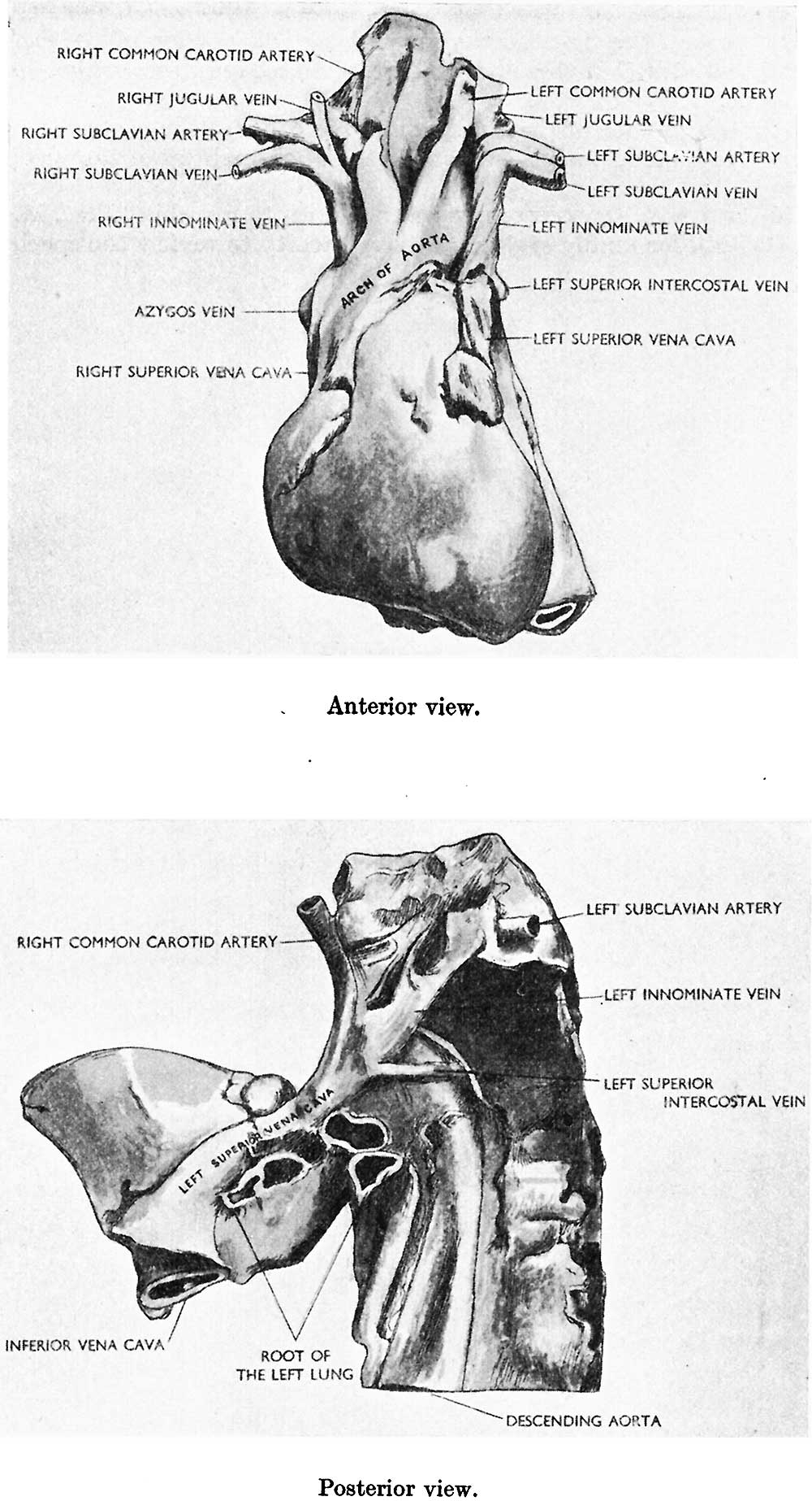
The two drawings—one of the anterior and the other of the posterior view of the heart and the great blood vessels—illustrate the abnormalities of the specimen.
Reference
Basu BN Persistent left superior vena cava, left duct of cuvier and left horn of the sinus venosus. (1932) J Anat. 66(2): 268–270. PMID 17104374
Cite this page: Hill, M.A. (2024, April 24) Embryology Basu1932 fig01.jpg. Retrieved from https://embryology.med.unsw.edu.au/embryology/index.php/File:Basu1932_fig01.jpg
- © Dr Mark Hill 2024, UNSW Embryology ISBN: 978 0 7334 2609 4 - UNSW CRICOS Provider Code No. 00098G
File history
Click on a date/time to view the file as it appeared at that time.
| Date/Time | Thumbnail | Dimensions | User | Comment | |
|---|---|---|---|---|---|
| current | 09:05, 29 November 2017 | | 1,000 × 1,846 (190 KB) | Z8600021 (talk | contribs) | |
| 09:05, 29 November 2017 | 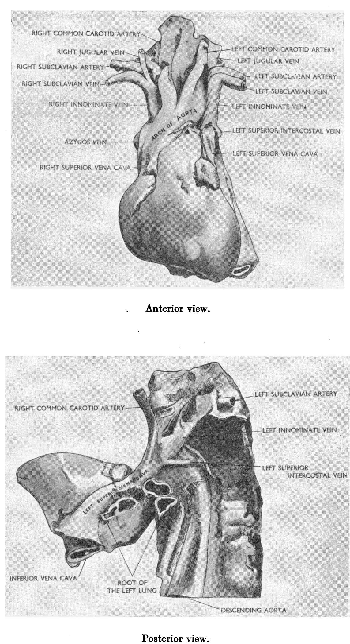 | 1,143 × 2,098 (406 KB) | Z8600021 (talk | contribs) |
You cannot overwrite this file.