File:Bardeen1908 fig1B.jpg
From Embryology
Size of this preview: 488 × 600 pixels. Other resolution: 1,496 × 1,838 pixels.
Original file (1,496 × 1,838 pixels, file size: 280 KB, MIME type: image/jpeg)
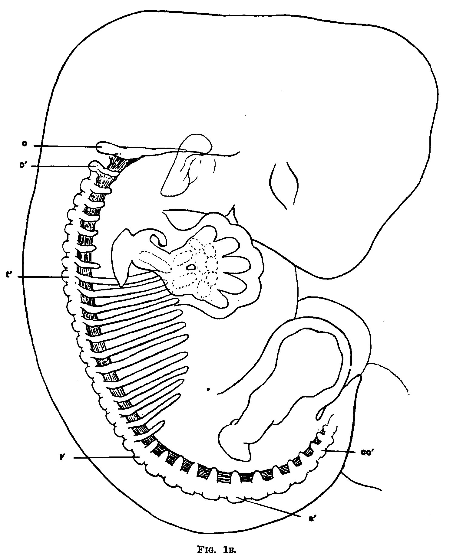
Fig. 1B. Diagram of the skeleton of an embryo 11 mm long and about five weeks old
- 0 - occipital plate.
- l' - first lumbar vertebra.
- c - flrst cervical vertebra.
- s' - first sacral vertebra.
- t’, that thoracic vertebra.
- co’, first coccygeal vertebra.
| Historic Disclaimer - information about historic embryology pages |
|---|
 |
- Links: Fig. 1A | Fig. 1B | Bardeen 1908 | Historic Embryology Papers
Reference
Bardeen CR. Vertebral regional determination in young human embryos. (1908) Amer. J Anat. 2: 99 - 105.
Cite this page: Hill, M.A. (2024, April 25) Embryology Bardeen1908 fig1B.jpg. Retrieved from https://embryology.med.unsw.edu.au/embryology/index.php/File:Bardeen1908_fig1B.jpg
- © Dr Mark Hill 2024, UNSW Embryology ISBN: 978 0 7334 2609 4 - UNSW CRICOS Provider Code No. 00098G
File history
Click on a date/time to view the file as it appeared at that time.
| Date/Time | Thumbnail | Dimensions | User | Comment | |
|---|---|---|---|---|---|
| current | 17:48, 28 October 2015 | | 1,496 × 1,838 (280 KB) | Z8600021 (talk | contribs) | |
| 17:48, 28 October 2015 |  | 1,496 × 2,058 (341 KB) | Z8600021 (talk | contribs) |
You cannot overwrite this file.
File usage
The following page uses this file: