File:Bardeen1908 fig1A.jpg
From Embryology
Size of this preview: 501 × 599 pixels. Other resolution: 836 × 1,000 pixels.
Original file (836 × 1,000 pixels, file size: 90 KB, MIME type: image/jpeg)
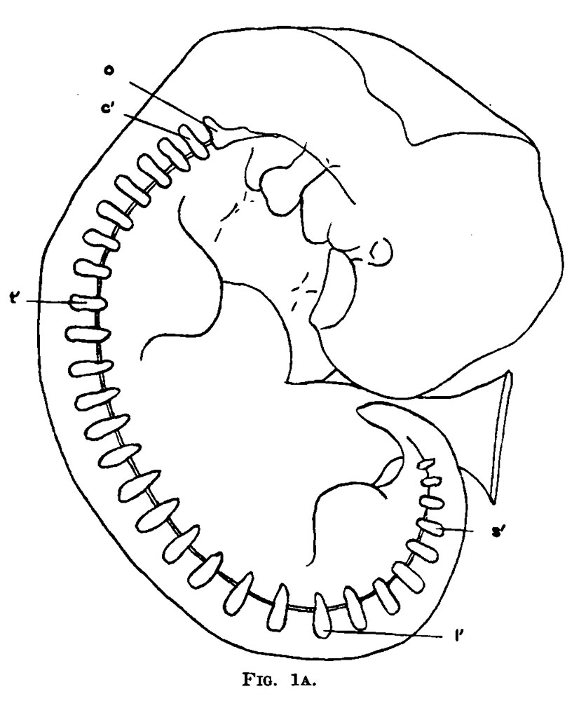
Fig. 1A. Diagram of the vertebral column of an embryo 7.5 mm long and about four weeks old
0, occipital plate. t’, first thoracic scleromere. c, first occipital scleromere. s‘, first sacral scleromere. l‘, first lumbar scleromere.
| Historic Disclaimer - information about historic embryology pages |
|---|
 |
- Links: Fig. 1A | Fig. 1B | Bardeen 1908 | Historic Embryology Papers
Reference
Bardeen CR. Vertebral regional determination in young human embryos. (1908) Amer. J Anat. 2: 99 - 105.
Cite this page: Hill, M.A. (2024, April 19) Embryology Bardeen1908 fig1A.jpg. Retrieved from https://embryology.med.unsw.edu.au/embryology/index.php/File:Bardeen1908_fig1A.jpg
- © Dr Mark Hill 2024, UNSW Embryology ISBN: 978 0 7334 2609 4 - UNSW CRICOS Provider Code No. 00098G
File history
Click on a date/time to view the file as it appeared at that time.
| Date/Time | Thumbnail | Dimensions | User | Comment | |
|---|---|---|---|---|---|
| current | 17:46, 28 October 2015 | | 836 × 1,000 (90 KB) | Z8600021 (talk | contribs) | |
| 17:45, 28 October 2015 | 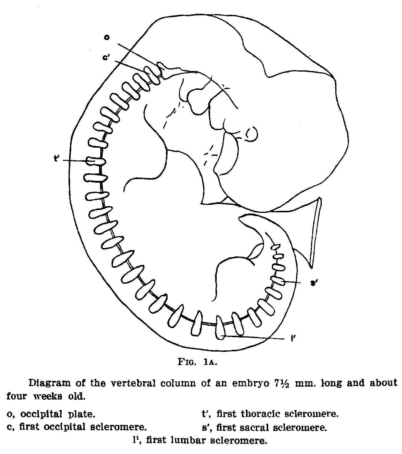 | 1,343 × 1,516 (198 KB) | Z8600021 (talk | contribs) |
You cannot overwrite this file.
File usage
The following page uses this file: