File:Bailey371.jpg
Original file (687 × 997 pixels, file size: 97 KB, MIME type: image/jpeg)
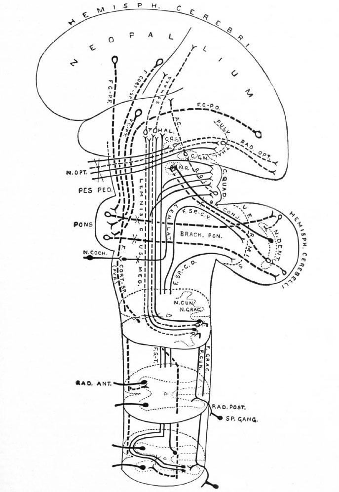
Fig. 371. Principal afferent and efferent suprasegmental pathways
(excepting the archipallial connections, the efferent connections of the mid- brain roof and the olivo-cerebellar connections)
Neopallial connections are indicated by broken lines. Intersegmental connections are omitted Some peripheral elements are indicated. Each neurone group (nucleus and fasciculus) is in dicated by one or several individual neurones. Decussations of tracts are indicated by an X
ac., Acoustic radiation, from medial gemculate body to temporal lobe; Z>r. conj., brachium conjunctivum (superior cerebellar peduncle); brack, pon., brachium ponds (middle cerebellar peduncle); b.q. i., brachium quadrigeminum inferias (a link in the cochlear pathway) ; c. g. I., lateral or external geniculate body; c. g. m., medial or internal geniculate body; c. quad., corpora quadrigemina; f.cort.-sp., cortico-spinal fasciculus (pyramidal tract);/. c. p.-f. frontal cortico-pontile fasciculus (from frontal lobe); f.c.-p.t., temporal cortico-pontile fasciculus (from temporal lobe); f.c.-p.o., occipital cortico-pontile fasciculus (from occipital lobe); f.ctm.f fasciculus cuneatus (column of Burdach); f.grac., fasciculus gracilis (column of Goll) ; /. s.-t., tract from cord to mid-brain roof and thalamus (sometimes included in Gowers* tract); f.sp.-c.d., dorsal spino-cerebellar fasciculus (tract of Flechsig); f.sp.-c.v., ventral spino-cerebellar fasciculus (tract of Gowers, location of cells in cord uncertain) ; lem. lot., lateral lemniscus or lateral fillet; lemniscus*-med., medial lemniscus or fillet (the part to the thalamus is mainly a neopallial acquisition); n.coch., cochlear nerve; n. cun., (terminal) nucleus of the column of Burdach; n.grac., nucleus of the column of Goll; n.dent., nucleus dentatus; n. opt., optic nerve; n.r., nucleus ruber (red nucleus); pes ped., pes pedunculi (crusta); pulv. thai., pulvinar thalami; pyr., pyramid; rod. ant., ventral spinal root; rod. post,. dorsal spinal root; rod. opt., optic radiation (from lateral geniculate body, and pulvinar (?), to calcarine region); somaes., bundles from thalamus to postcentral region of neopallium; s p. gang., spinal ganglion; ihal., thalamus.
- Text-Book of Embryology: Germ cells | Maturation | Fertilization | Amphioxus | Frog | Chick | Mammalian | External body form | Connective tissues and skeletal | Vascular | Muscular | Alimentary tube and organs | Respiratory | Coelom, Diaphragm and Mesenteries | Urogenital | Integumentary | Nervous System | Special Sense | Foetal Membranes | Teratogenesis | Gallery of All Figures
| Historic Disclaimer - information about historic embryology pages |
|---|
 |
Reference
Bailey FR. and Miller AM. Text-Book of Embryology (1921) New York: William Wood and Co.
Cite this page: Hill, M.A. (2024, April 24) Embryology Bailey371.jpg. Retrieved from https://embryology.med.unsw.edu.au/embryology/index.php/File:Bailey371.jpg
- © Dr Mark Hill 2024, UNSW Embryology ISBN: 978 0 7334 2609 4 - UNSW CRICOS Provider Code No. 00098G
File history
Click on a date/time to view the file as it appeared at that time.
| Date/Time | Thumbnail | Dimensions | User | Comment | |
|---|---|---|---|---|---|
| current | 23:49, 29 January 2011 | | 687 × 997 (97 KB) | S8600021 (talk | contribs) | {{Template:Bailey 1921 Figures}} Category:Neural |
You cannot overwrite this file.
File usage
The following 4 pages use this file: