File:Bailey366.jpg
Original file (558 × 633 pixels, file size: 97 KB, MIME type: image/jpeg)
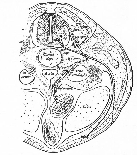
Fig. 366. Transverse section through the body of a typical Vertebrate
Showing the peripheral (segmental) nervous apparatus.
Froriep.
Small dots, afferent visceral neurones; coarse dots, afferent somatic neurones; dashes, efferent visceral (ventral root and sympathetic) neurones; lines, efferent somatic neurones.
Darm, gut; Ggl. spin., spinal ganglion; Ggl. vert., vertebral sympathetic ganglion; Ggl. mesent., mesenteric sympathetic ganglion. The peripheral sympathetic ganglionic plexuses (Auerbach and Meissner) are not shown. Muse., muscle; Rad. dors., dorsal root; Rad. vent., ventral root; R. comm., white ramus communicans.
Two sympathetic neurones are represented as intercalated in the visceral efferent pathway. It doubtful if there should be more than one.
- Another important differentiation arises apparently from the important physiological difference in general character between the activities of what may be termed the internal (visceral or splanchnic) and the external (somatic) structures. Internal activities are to a certain extent independent of activities which have to do more with the reactions of the organism to the external world, and consequently their nervous mechanisms have a more or less independent character, forming what is often called the autonomic (sympathetic) system. This independence is exhibited structurally by the intercalation in the peripheral pathway of additional neurones, whose bodies form visceral ganglia connected in various ways among themselves and probably having their own reflex arcs or plexuses. These ganglia are nevertheless to some extent under the control of the efferent neurones of the central nervous system, some of which send their axones to such ganglia (Fig. 366). There are thus in the central nervous system two categories of efferent peripheral neurones, those innervating visceral structures "via sympathetic ganglia and those innervating somatic structures. The b'odies of the somatic efferent neurones are located in the ventral gray matter of the nerve tube, while the bodies of the splanchnic efferent neurones are believed to occupy more central and lateral positions in the lower half of the gray matter of the neural tube (Fig. 366). It is uncertain whether there are similar afferent splanchnic neurones in the sympathetic ganglia, and thus distinct from those in the spinal ganglia, or whether these all lie in the spinal ganglia and are consequently not fully differentiated from the somatic afferent neurones.
- Text-Book of Embryology: Germ cells | Maturation | Fertilization | Amphioxus | Frog | Chick | Mammalian | External body form | Connective tissues and skeletal | Vascular | Muscular | Alimentary tube and organs | Respiratory | Coelom, Diaphragm and Mesenteries | Urogenital | Integumentary | Nervous System | Special Sense | Foetal Membranes | Teratogenesis | Gallery of All Figures
| Historic Disclaimer - information about historic embryology pages |
|---|
 |
Reference
Bailey FR. and Miller AM. Text-Book of Embryology (1921) New York: William Wood and Co.
Cite this page: Hill, M.A. (2024, April 25) Embryology Bailey366.jpg. Retrieved from https://embryology.med.unsw.edu.au/embryology/index.php/File:Bailey366.jpg
- © Dr Mark Hill 2024, UNSW Embryology ISBN: 978 0 7334 2609 4 - UNSW CRICOS Provider Code No. 00098G
File history
Click on a date/time to view the file as it appeared at that time.
| Date/Time | Thumbnail | Dimensions | User | Comment | |
|---|---|---|---|---|---|
| current | 16:41, 29 January 2011 | | 558 × 633 (97 KB) | S8600021 (talk | contribs) | ==Fig. 366. Transverse section through the body of a typical Vertebrate== Showing the peripheral (segmental) nervous apparatus. Froriep. Small dots, afferent visceral neurones; coarse dots, afferent somatic neurones; dashes, efferent visceral (ventra |
You cannot overwrite this file.