File:Bailey356.jpg
Bailey356.jpg (767 × 533 pixels, file size: 103 KB, MIME type: image/jpeg)
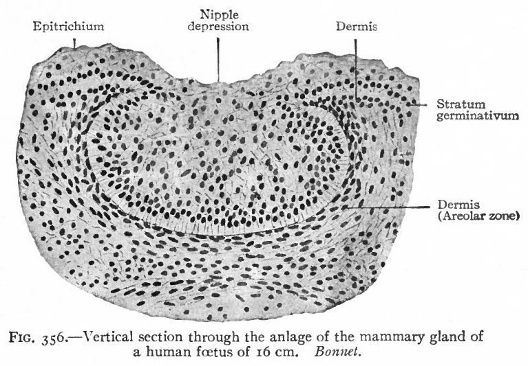
Fig. 356. Vertical section through the anlage of the mammary gland of a human foetus of 16 cm
Bonnet.
In embryos of six to seven mm., or even less, a thickening of the epidermis occurs in a narrow zone along the ventro-lateral surface of the body (Strahl). In embryos of 15 mm. this thickening, known as the milk ridge, extends from the upper extremity to the inguinal region (Kallius, Schmidt). Later the caudal end of the ridge disappears, while the cephalic portion becomes more prominent. The further history of the ridge has not been traced, but in embryos considerably older the anlage of each gland is a circular thickening of the epidermis in the thoracic region, projecting into the underlying dermis. It seems most probable that this local thickening represents a portion of the original ridge, the remainder having disappeared.
Later the central cells of the epidermal mass become cornified and are cast off, leaving a depression in the skin (Fig. 356). In embryos of 250 mm. a number of solid secondary buds have grown out (Fig. 357). These resemble the anlagen of the sweat glands, to which they are generally considered as closely allied (Hertwig, Wiedersheim and others), and represent the excretory ducts. Continued evaginations from the terminal parts of the excretory ducts form the lobular ducts and acini. The acini, however, are scarcely demonstrable in the male, and not even in the female until pregnancy. Lumina appear by a separation and breaking down of the central cells of the ducts and acini, the peripheral cells remaining as their lining.
- Text-Book of Embryology: Germ cells | Maturation | Fertilization | Amphioxus | Frog | Chick | Mammalian | External body form | Connective tissues and skeletal | Vascular | Muscular | Alimentary tube and organs | Respiratory | Coelom, Diaphragm and Mesenteries | Urogenital | Integumentary | Nervous System | Special Sense | Foetal Membranes | Teratogenesis | Gallery of All Figures
| Historic Disclaimer - information about historic embryology pages |
|---|
 |
Reference
Bailey FR. and Miller AM. Text-Book of Embryology (1921) New York: William Wood and Co.
Cite this page: Hill, M.A. (2024, April 19) Embryology Bailey356.jpg. Retrieved from https://embryology.med.unsw.edu.au/embryology/index.php/File:Bailey356.jpg
- © Dr Mark Hill 2024, UNSW Embryology ISBN: 978 0 7334 2609 4 - UNSW CRICOS Provider Code No. 00098G
File history
Click on a date/time to view the file as it appeared at that time.
| Date/Time | Thumbnail | Dimensions | User | Comment | |
|---|---|---|---|---|---|
| current | 16:01, 29 January 2011 | | 767 × 533 (103 KB) | S8600021 (talk | contribs) | ==Fig. 356. Vertical section through the anlage of the mammary gland of a human foetus of 16 cm== Bonnet. {{Template:Bailey 1921 Figures}} Category:Human Category:Integumentary Category:Mammary Category:Fetus |
You cannot overwrite this file.
File usage
The following 3 pages use this file: